. 2016 Jun 29;17(1):141.
doi: 10.1186/s13059-016-1001-5.

In vivo genome-wide profiling reveals a tissue-specific role for 5-formylcytosine

Affiliations

In vivo genome-wide profiling reveals a tissue-specific role for 5-formylcytosine

Mario Iurlaro et al. Genome Biol. .

Abstract

Background: Genome-wide methylation of cytosine can be modulated in the presence of TET and thymine DNA glycosylase (TDG) enzymes. TET is able to oxidise 5-methylcytosine (5mC) to 5-hydroxymethylcytosine (5hmC), 5-formylcytosine (5fC) and 5-carboxylcytosine (5caC). TDG can excise the oxidative products 5fC and 5caC, initiating base excision repair. These modified bases are stable and detectable in the genome, suggesting that they could have epigenetic functions in their own right. However, functional investigation of the genome-wide distribution of 5fC has been restricted to cell culture-based systems, while its in vivo profile remains unknown.

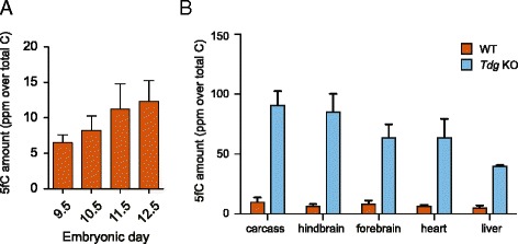
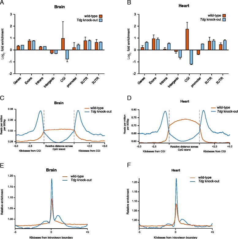
Results: Here, we describe the first analysis of the in vivo genome-wide profile of 5fC across a range of tissues from both wild-type and Tdg-deficient E11.5 mouse embryos. Changes in the formylation profile of cytosine upon depletion of TDG suggest TET/TDG-mediated active demethylation occurs preferentially at intron-exon boundaries and reveals a major role for TDG in shaping 5fC distribution at CpG islands. Moreover, we find that active enhancer regions specifically exhibit high levels of 5fC, resulting in characteristic tissue-diagnostic patterns, which suggest a role in embryonic development.

Conclusions: The tissue-specific distribution of 5fC can be regulated by the collective contribution of TET-mediated oxidation and excision by TDG. The in vivo profile of 5fC during embryonic development resembles that of embryonic stem cells, sharing key features including enrichment of 5fC in enhancer and intragenic regions. Additionally, by investigating mouse embryo 5fC profiles in a tissue-specific manner, we identify targeted enrichment at active enhancers involved in tissue development.


Figures

Fig. 1
Fig. 1
Genomic 5fC is heterogeneous in early embryos. a LC/MS quantification of genomic 5fC levels in mid-gestation embryos. Displayed is the average of at least three biological replicates with standard deviation. Results are expressed as ppm (part per million over total of cytosines). b LC/MS quantification of genomic 5fC levels in dissected tissues from WT (brown) and Tdg knockout (light blue) E11.5 embryos. Displayed is the average of three biological replicates with standard deviation. Results are expressed as ppm (part per million over total of cytosines)
Fig. 2
Fig. 2
Genome-wide profiling of 5fC in vivo. a, b Enrichment of 5fC peaks over functional genomic features in E11.5 hindbrain and heart, respectively. Log2 fold enrichment was calculated for each replicate individually using 10,000 randomisations in a simulation procedure implemented with the GAT [21]. Plotted is the average enrichment with standard deviation. c, d Trend plot showing 5fC profile over CpG islands (+/− 5 kb) in the hindbrain and heart, respectively. e, f Trend plot showing 5fC profile over exon/intron boundaries (+/− 5 kb) in the hindbrain and heart, respectively
Fig. 3
Fig. 3
5fC is enriched at active enhancers in vivo. a Screenshot of a genomic region on chromosome 2 exemplifying the striking overlap of the 5fC signal with regions marked by H3K4me1 (orange boxes) and H3K27ac (purple boxes), as calculated in [27]. b Fold enrichment of 5fC peaks in hindbrain and heart from WT and Tdg null embryos over regions marked by H3K4me1, H3K4me3 and H3K27ac in the brain and heart of E14.5 embryos, respectively. Log2 fold enrichment was calculated for each replicate individually using 10,000 randomisations in a simulation procedure implemented with the GAT. Plotted is the average enrichment with standard deviation. c Comparison of 5fC peak enrichment in the hindbrain in regions marked by different histone modifications in the E14.5 mouse brain
Fig. 4
Fig. 4
Tissue-specific 5fC formation at developmental enhancers. a Enrichment of 5fC peaks in the different tissues over tissue-specific enhancer regions. Log2 fold enrichment was calculated for each replicate individually using 10,000 randomisations in a simulation procedure implemented with the GAT. Plotted is the average enrichment with standard deviation. b Comparison of 5fC peaks detected specifically in the hindbrain and heart samples. Gene Ontology analysis was performed on genes in proximity (1 kb cutoff) of hindbrain-specific and heart-specific peaks

References

    1. Tahiliani M, Koh KP, Shen Y, Pastor WA, Bandukwala H, Brudno Y, et al. Conversion of 5-methylcytosine to 5-hydroxymethylcytosine in mammalian DNA by MLL partner TET1. Science. 2009;324:930–5. doi: 10.1126/science.1170116. - DOI - PMC - PubMed
    1. Ito S, D'Alessio AC, Taranova OV, Hong K, Sowers LC, Zhang Y. Role of Tet proteins in 5mC to 5hmC conversion, ES-cell self-renewal and inner cell mass specification. Nature. 2010;466:1129–33. doi: 10.1038/nature09303. - DOI - PMC - PubMed
    1. Kriaucionis S, Heintz N. The nuclear DNA base 5-hydroxymethylcytosine is present in Purkinje neurons and the brain. Science. 2009;324:929–30. doi: 10.1126/science.1169786. - DOI - PMC - PubMed
    1. Ito S, Shen L, Dai Q, Wu SC, Collins LB, Swenberg JA, et al. Tet proteins can convert 5-methylcytosine to 5-formylcytosine and 5-carboxylcytosine. Science. 2011;333:1300–3. doi: 10.1126/science.1210597. - DOI - PMC - PubMed
    1. He YF, Li BZ, Li Z, Liu P, Wang Y, Tang Q, et al. Tet-mediated formation of 5-carboxylcytosine and its excision by TDG in mammalian DNA. Science. 2011;333:1303–7. doi: 10.1126/science.1210944. - DOI - PMC - PubMed

Publication types