. 2016 Jun 30:7:12038.
doi: 10.1038/ncomms12038.

Coronary vasculature patterning requires a novel endothelial ErbB2 holoreceptor

Affiliations

Coronary vasculature patterning requires a novel endothelial ErbB2 holoreceptor

Haig Aghajanian et al. Nat Commun. .

Abstract

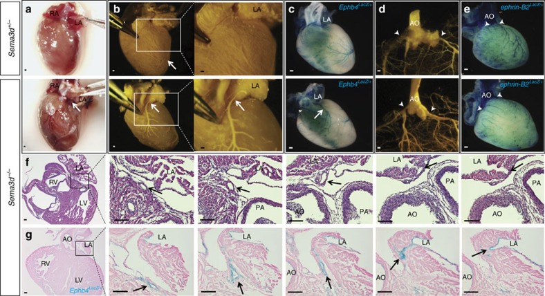
Organogenesis and regeneration require coordination of cellular proliferation, regulated in part by secreted growth factors and cognate receptors, with tissue nutrient supply provided by expansion and patterning of blood vessels. Here we reveal unexpected combinatorial integration of a growth factor co-receptor with a heterodimeric partner and ligand known to regulate angiogenesis and vascular patterning. We show that ErbB2, which can mediate epidermal growth factor (EGF) and neuregulin signalling in multiple tissues, is unexpectedly expressed by endothelial cells where it partners with neuropilin 1 (Nrp1) to form a functional receptor for the vascular guidance molecule semaphorin 3d (Sema3d). Loss of Sema3d leads to improper patterning of the coronary veins, a phenotype recapitulated by endothelial loss of ErbB2. These findings have implications for possible cardiovascular side-effects of anti-ErbB2 therapies commonly used for cancer, and provide an example of integration at the molecular level of pathways involved in tissue growth and vascular patterning.


Figures

Figure 1
Figure 1. Coronary veins abnormally connect to the left atrium in Sema3d/ hearts.
(a) Left anterior oblique view of adult Sema3d+/− (top) and Sema3d−/− (bottom) hearts. The anterior interventricular vein (arrow) can be seen connecting to the left atrium in the Sema3d−/− heart. (b) Mircrofil perfusion of 3 week old hearts from Sema3d+/− (top) and Sema3d/ (bottom) mice. (c) Whole-mount X-gal staining of Sema3d+/−;Ephb4LacZ/+ (top) and Sema3d−/−;Ephb4LacZ/+ (bottom) postnatal hearts. (d) Cleared Sema3d+/− (top) and Sema3d−/− (bottom) postnatal hearts perfused with Microfil to visualize the origin of the coronary arteries (arrowheads). (e) Whole-mount X-gal staining of Sema3d+/−;ephrin-B2LacZ/+ (top) and Sema3d−/−;ephrin-B2LacZ/+ (bottom) postnatal hearts verifying arterial identity of the coronary vessels originating from the aorta (arrowheads). (f) Photomicrographs of H&E stained frontal sections from a Sema3d−/− heart following a coronary vein (arrows) from the left anterior AV groove connecting with the left atrial lumen. (g) Photomicrographs of eosin and X-gal stained frontal sections from a postnatal Sema3d−/−;Ephb4LacZ/+ heart following a coronary vein (arrows) from the left anterior AV groove connecting with the left atrial lumen. AO, aorta; LA, left atrium; LV, left ventricle; PA, pulmonary artery; RV, right ventricle. Scale bars, 100 μm.
Figure 2
Figure 2. Sema3d expression excludes endothelium from the AV groove.
(a,b) X-gal and eosin staining of Sema3dGFPCre/+;Ephb4LacZ/+ (a) and Sema3dGFPCre/GFPCre;Ephb4LacZ/+ (b) E15.5 heart frontal sections. Numerous LacZ positive (venous) cells can be observed in the left anterior AV junction of the Sema3d nulls (b; arrow) but not in the control (a). (c,d) Immunofluorescence for the endothelial marker eNOS (red) and GFP (green) of E15.5 Sema3dGFPCre/+;Ephb4LacZ/+ (c) and Sema3dGFPCre/GFPCre;Ephb4LacZ/+ (d) heart frontal sections. The GFP expressing left anterior AV groove (arrow) is mostly avascular in the control (c) but penetrated by numerous eNOS-positive (red) vessels in the Sema3d null (d). (e) Model depicting the role of Sema3d in coronary venous connection. In wild type mice, Sema3d is expressed in the left anterior AV groove and repels venous endothelium from abnormally forming connections with the left atrium (left), but in the absence of Sema3d the coronary anterior interventricular vein (AIV) abnormally connects to the LV (right). LA, left atrium; LV, left ventricle; RA, right atrium; RV, right ventricle. Scale bars, 100 μm.
Figure 3
Figure 3. Sema3d signals via ErbB2 in endothelial cells.
(a) Western blot for phospho-ErbB2 (Y1248) from HUVECs incubated with increasing concentrations of Sema3d for 30 min. (b) Transwell migration assay showing the percentage HUVECs that migrated through a porous membrane in the presence of Sema3d or Sema3e when compared to vehicle control in the presence of a control siRNA (left) or ErbB2 siRNA (right). ***P<0.001, *P<0.05, NS, not significant (one-way ANOVA between groups P<0.001 for both groups; post-hoc multiple comparisons, Tukey's test) (c) Western blot for phospho-Akt (S473) and phospho-ErbB2 (Y1248) from HUVECs incubated with increasing concentrations of Sema3d for 5 min. (d) Western blot for ErbB2 and phospho-Akt (S473) of HUVECs in the presence of control or ErbB2 siRNA treated with Sema3d (10 nM) for 5 min. (e) Quantification of ErbB2 knockdown and Akt phosphorylation from (d). **P<0.01, *P<0.05 (Student's t-test, n=3).
Figure 4
Figure 4. ErbB2 is expressed by coronary venous endothelium.
(a) Immunofluorescence of a frontal section of an E14.5 heart for the endothelial marker eNOS (red) and ErbB2 (green). ErbB2 and eNOS co-localize to vessels in the antrioventricular junction (arrowheads). (b) Frontal section of an E17.5 heart co-stained for eNOS (red) and ErbB2 (green) demonstrating co-localization in subepicardial vessels (white arrowheads) but not in intramyocardial vessels (yellow arrowheads). (c) Phospho-ErbB2 (Y1248) immunofluorescence from frontal sections of E14.5 Sema3d+/− and Sema3d−/− hearts. ErbB2 phosphorylation is reduced in the Sema3d−/− hearts (arrowheads). (d) Photomicrographs of H&E stained frontal sections from a postnatal ErbB2fl/−;Tie2-Cre heart tracing a vessel from the left anterior AV groove to the left atrial lumen (arrows). LA, left atrium; LV, left ventricle. Scale bars, 100 μm.
Figure 5
Figure 5. ErbB2 and Nrp1 interact to form a Sema3d receptor.
(a) Representative photomicrographs of Cos-7 cells expressing indicated receptors after binding and enzymatic detection of alkaline phosphatase tagged Sema3d protein. (b) Co-immunoprecipitation of Nrp1 and ErbB2 in the presence or absence of Sema3d (10 nM). (c) Graphical representation of ErbB2 deletion constructs (top). Co-immunoprecipitation of Nrp1 and either full length or truncated ErbB2. (d) Western blot and quantification for phospho-ErbB2 (Y1248) from HUVECs treated with a Nrp1 or control siRNA, with or without Sema3d for 5 min. *P<0.05 (Student's t-test, n=4). (e) Immunofluorescence of E15.5 hearts shows that ErbB2 (green) and Nrp1 (red) co-localize in coronary vessels. (f) Model for Sema3d signalling to coronary endothelial cells. Sema3d binds to the Nrp1 subunit of a Nrp1/ErbB2 complex on endothelial cells. This results in the phosphorylation and activation of ErbB2 and subsequent downstream signalling, including phosphorylation of Akt. ECD, extracellular domain; ICD, intracellular domain; TM, transmembrane domain. Scale bars: a,100 μm; e, 10 μm.

References

    1. Angelini P. Coronary artery anomalies: an entity in search of an identity. Circulation 115, 1296–1305 (2007). - PubMed
    1. Hutchins G. M., Kessler-Hanna A. & Moore G. W. Development of the coronary arteries in the embryonic human heart. Circulation 77, 1250–1257 (1988). - PubMed
    1. Bogers A. J., Gittenberger-de Groot A. C., Poelmann R. E., Peault B. M. & Huysmans H. A. Development of the origin of the coronary arteries, a matter of ingrowth or outgrowth? Anat. Embryol. (Berl) 180, 437–441 (1989). - PubMed
    1. Katz T. C. et al. Distinct compartments of the proepicardial organ give rise to coronary vascular endothelial cells. Dev. Cell 22, 639–650 (2012). - PMC - PubMed
    1. Red-Horse K., Ueno H., Weissman I. L. & Krasnow M. A. Coronary arteries form by developmental reprogramming of venous cells. Nature 464, 549–553 (2010). - PMC - PubMed

Publication types