. 2016 Jun 29;11(1):47.
doi: 10.1186/s13024-016-0109-0.

Acetylated tau destabilizes the cytoskeleton in the axon initial segment and is mislocalized to the somatodendritic compartment

Affiliations

Acetylated tau destabilizes the cytoskeleton in the axon initial segment and is mislocalized to the somatodendritic compartment

Peter Dongmin Sohn et al. Mol Neurodegener. .

Abstract

Background: Neurons are highly polarized cells in which asymmetric axonal-dendritic distribution of proteins is crucial for neuronal function. Loss of polarized distribution of the axonal protein tau is an early sign of Alzheimer's disease (AD) and other neurodegenerative disorders. The cytoskeletal network in the axon initial segment (AIS) forms a barrier between the axon and the somatodentritic compartment, contributing to axonal retention of tau. Although perturbation of the AIS cytoskeleton has been implicated in neurological disorders, the molecular triggers and functional consequence of AIS perturbation are incompletely understood.

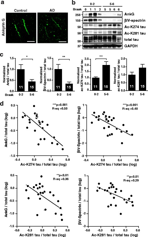
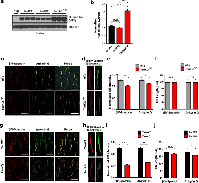
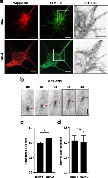
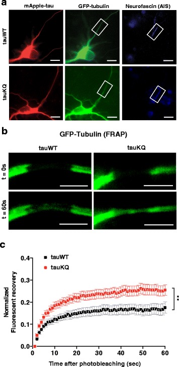
Results: Here we report that tau acetylation and consequent destabilization of the AIS cytoskeleton promote the somatodendritic mislocalization of tau. AIS cytoskeletal proteins, including ankyrin G and βIV-spectrin, were downregulated in AD brains and negatively correlated with an increase in tau acetylated at K274 and K281. AIS proteins were also diminished in transgenic mice expressing tauK274/281Q, a tau mutant that mimics K274 and K281 acetylation. In primary neuronal cultures, the tauK274/281Q mutant caused hyperdynamic microtubules (MTs) in the AIS, shown by live-imaging of MT mobility and fluorescence recovery after photobleaching. Using photoconvertible tau constructs, we found that axonal tauK274/281Q was missorted into the somatodendritic compartment. Stabilizing MTs with epothilone D to restore the cytoskeletal barrier in the AIS prevented tau mislocalization in primary neuronal cultures.

Conclusions: Together, these findings demonstrate that tau acetylation contributes to the pathogenesis of neurodegenerative disease by compromising the cytoskeletal sorting machinery in the AIS.

Keywords: Alzheimer’s disease; Axon initial segment; Neuronal cytoskeleton; Neuronal polarity; Tau acetylation.


Figures

Fig. 1
Fig. 1
Levels of AIS cytoskeletal proteins are downregulated in human AD brains and correlate negatively with ac-K274 and ac-K281 tau levels. a Representative images of AnkG immunostaining in human control and AD brains. Scale bars, 10 μm. b, c Representative western blots and quantification of levels of AnkG, βIV-spectrin, ac-K274 tau, and ac-K281 tau in human AD brains. n = 11–15 samples/group. *p < 0.05, **p < 0.01, unpaired t test. d Correlation analyses between AnkG or βIV-spectrin and ac-K274 or ac-K281 tau. Pearson correlation analyses after natural log transformation. Values are mean ± SEM (c)
Fig. 2
Fig. 2
Expression of human K274/281Q tau in transgenic mice destabilizes the AIS cytoskeleton. a, b Levels of expression of human tau in the cortex of tauWT and tauKQ mice. n = 7-8 mice/group. ***p < 0.001, one-way ANOVA Bonferonni post-hoc analyses. c Representative images of βIV-spectrin and AnkG immunostaining in the somatosensory cortex of 2 month-old nontransgenic and tauKQhigh mice. Scale bars, 20 μm. d-f Representative images and quantification of the intensity and length of βIV-spectrin and AnkG immunostaining in the somatosensory cortex of 2 month-old non-transgenic and tauKQhigh mice. n = 119–132 cells from 6 mice/group. *p < 0.05, **p < 0.01, mixed-model linear regression analyses. Scale bars, 5 μm. g Representative images of βIV-spectrin and AnkG immunostaining in the somatosensory cortex of 10-12 month-old tauWT and tauKQ mice. Scale bars, 20 μm. h-j Representative images and quantification of intensity and length of βIV-spectrin and AnkG immunostaining in the somatosensory cortex of 10-12 month-old tauWT and tauKQ mice. n = 109-169 cells from 8 mice/group. *p < 0.05, **p < 0.01, mixed model linear regression analyses. Scale bars, 5 μm. Values are mean ± SEM
Fig. 3
Fig. 3
K274/281Q tau increases MT dynamics in primary neurons. a–d Measuring MT dynamics with GFP-EB3 in rat primary neurons. a Representative images of HeLa cells co-transfected with mApple-tauWT or -tauKQ and GFP-EB3. Panels on the right show enlarged images of GFP-EB3 comets within the white boxes. Scale bars, 5 μm. b Movement of an individual GFP-EB3 comet (arrowheads) Scale bar, 2 μm. c, d Quantification of GFP-EB3 movement rate and tau levels in rat primary neurons co-transfected with GFP-EB3 and mApple-tau. n = 11–13 cells/group from two independent experiments. *p < 0.05, one-way ANOVA with Tukey’s post hoc test. Values are mean ± SEM
Fig. 4
Fig. 4
K274/281Q tau increases tubulin dynamics in the AIS. a Representative images of a rat primary neuron co-transfected with mApple-tauWT or -tauKQ and GFP-tubulin. White boxes indicate the portion of the AIS (revealed by anti-neurofascin) where GFP-tubulin was photobleached. b Representative images of photobleaching and fluorescence recovery of GFP-tubulin in a ~5-μm portion of the AIS in rat primary neurons transfected with mApple-tauWT or -tauKQ. c Quantification of FRAP of GFP-tubulin in the AIS of rat primary neurons co-transfected with mApple-tauWT or -tauKQ and GFP-tubulin. n = 25–26 cells/group from three independent experiments. **p < 0.01, mixed model linear regression analysis. Values are mean ± SEM. Scale bars, 5 μm
Fig. 5
Fig. 5
Stabilization of MTs reduces somatodendritic mislocalization of K274/281Q tau. a–d Photoconversion of mEOS2-tau and its movement in rat primary neurons. a Schematic diagram of photoconversion of mEOS2-tau in the AIS and monitoring its movement toward the somatodendritic compartment and distal axon. b Time-lapse live images of mEOS2-tauWT, -tauKQ, and -tauKR before and after photoconversion in an axon segment ~30 μm from the AIS. The last row represents photoconversion of mEOS2-tauKQ after EpoD treatment (20 nM). White circles indicate the somatodendritic compartment. Scale bars, 10 μm. c, d Quantification of fluorescent intensity in the somatodendritic compartment (c) and distal axon (d) for 30 min after photoconversion of mEOS2-tau in rat primary neurons. n = 8–21 cells/group from three to nine independent experiments. *p < 0.05, **p < 0.01, mixed model linear regression analyses. Values are mean ± SEM

Similar articles

Cited by

References

    1. Ittner LM, Gotz J. Amyloid-beta and tau--a toxic pas de deux in Alzheimer’s disease. Nat Rev Neurosci. 2011;12:65–72. doi: 10.1038/nrn2967. - DOI - PubMed
    1. Zempel H, Mandelkow E. Lost after translation: missorting of Tau protein and consequences for Alzheimer disease. Trends Neurosci. 2014;37:721–732. doi: 10.1016/j.tins.2014.08.004. - DOI - PubMed
    1. Grundke-Iqbal I, Iqbal K, Tung YC, Quinlan M, Wisniewski HM, Binder LI. Abnormal phosphorylation of the microtubule-associated protein tau (tau) in Alzheimer cytoskeletal pathology. Proc Natl Acad Sci U S A. 1986;83:4913–4917. doi: 10.1073/pnas.83.13.4913. - DOI - PMC - PubMed
    1. Kosik KS, Joachim CL, Selkoe DJ. Microtubule-associated protein tau (tau) is a major antigenic component of paired helical filaments in Alzheimer disease. Proc Natl Acad Sci U S A. 1986;83:4044–4048. doi: 10.1073/pnas.83.11.4044. - DOI - PMC - PubMed
    1. Kowall NW, Kosik KS. Axonal disruption and aberrant localization of tau protein characterize the neuropil pathology of Alzheimer’s disease. Ann Neurol. 1987;22:639–643. doi: 10.1002/ana.410220514. - DOI - PubMed

Publication types

MeSH terms