. 2016 Jun 30:6:28916.
doi: 10.1038/srep28916.

Multiple cone pathways are involved in photic regulation of retinal dopamine

Affiliations

Multiple cone pathways are involved in photic regulation of retinal dopamine

Sheng-Nan Qiao et al. Sci Rep. .

Abstract

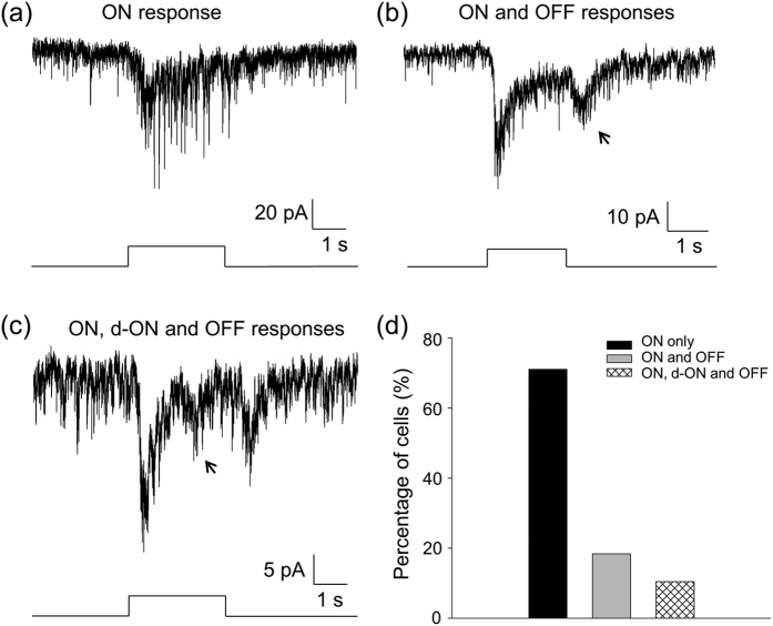
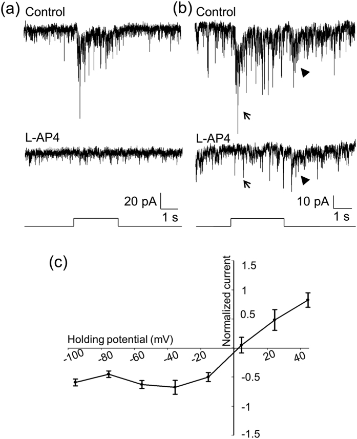
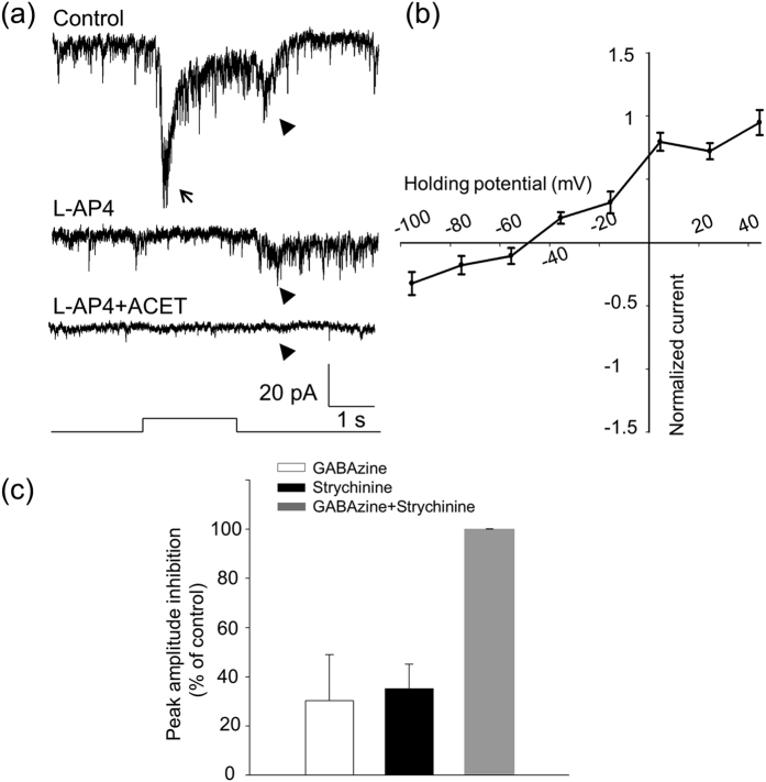
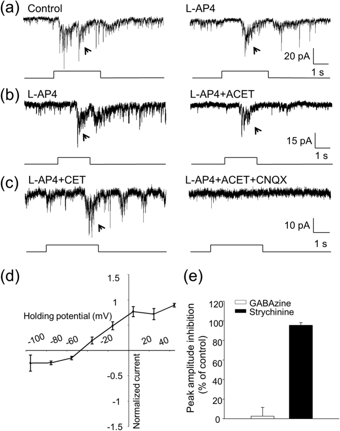
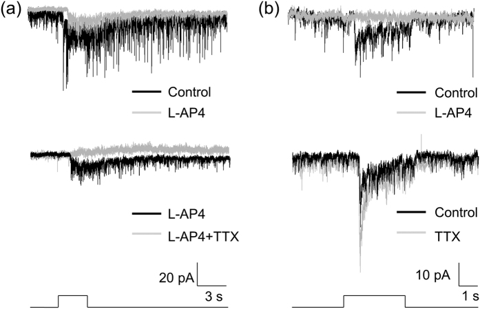
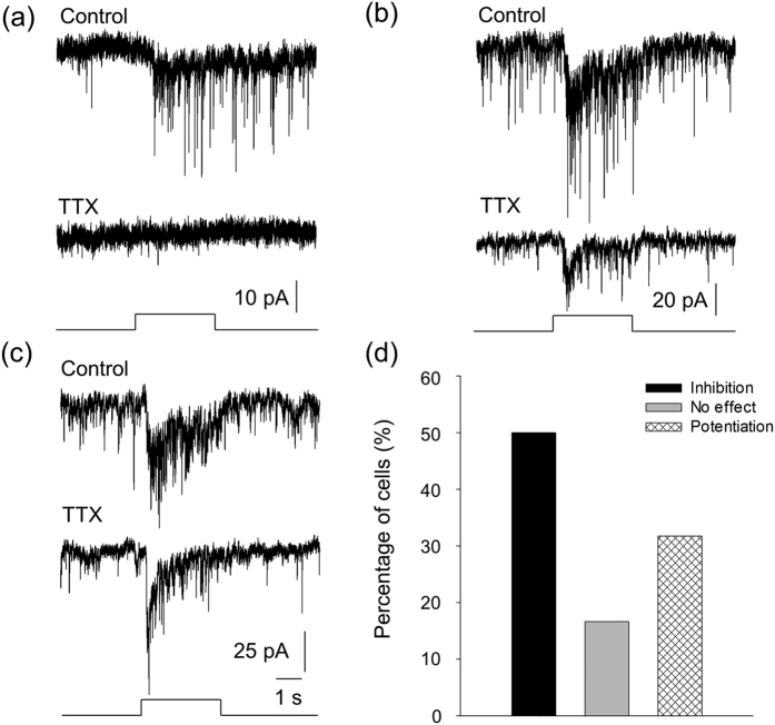
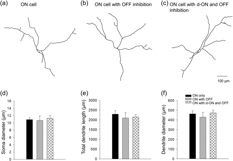
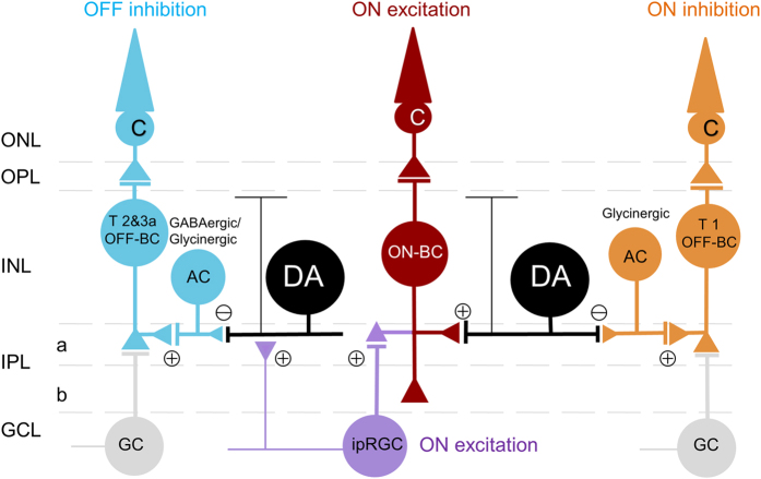
Dopamine is a key neurotransmitter in the retina and plays a central role in the light adaptive processes of the visual system. The sole source of retinal dopamine is dopaminergic amacrine cells (DACs). We and others have previously demonstrated that DACs are activated by rods, cones, and intrinsically photosensitive retinal ganglion cells (ipRGCs) upon illumination. However, it is still not clear how each class of photosensitive cells generates light responses in DACs. We genetically isolated cone function in mice to specifically examine the cone-mediated responses of DACs and their neural pathways. In addition to the reported excitatory input to DACs from light-increment (ON) bipolar cells, we found that cones alternatively signal to DACs via a retrograde signalling pathway from ipRGCs. Cones also produce ON and light-decrement (OFF) inhibitory responses in DACs, which are mediated by other amacrine cells, likely driven by type 1 and type 2/3a OFF bipolar cells, respectively. Dye injections indicated that DACs had similar morphological profiles with or without ON/OFF inhibition. Our data demonstrate that cones utilize specific parallel excitatory and inhibitory circuits to modulate DAC activity and efficiently regulate dopamine release and the light-adaptive state of the retina.


Figures

Figure 1
Figure 1. Cones generate three classes of light-evoked responses in DACs.
Whole-cell voltage-clamp recordings (Vhold = −65.5 mV) were made on DACs in flat-mount retinas isolated from cone-function-only mice. (a) A typical DAC exhibited a light-evoked inward current immediately after light onset (ON response). (b) In contrast to the cell in (a), this cell had an additional light-evoked inward current at light offset (OFF response, indicated by an arrow). (c) A typical cell displayed an inward current at ~1 s after light onset (d-ON response, arrow) between the ON and OFF responses. Stimulation bar under each trace shows timing of a 470-nm light pulse. Light stimulus duration was 3 s for (a) and (c) and 2 s for (b). (d) Of a total of 86 cells tested, 68.6% only had ON responses (black bar), 20.9% exhibited ON and OFF responses (grey bar), and 10.5% had all three classes of light responses (striped bar).
Figure 2
Figure 2. L-AP4 blocks ON responses of DACs that have an excitatory reversal potential.
(a) The ON response of a DAC (top trace) was completely blocked by L-AP4 (bottom trace). (b) This cell had ON and OFF responses (top trace). The bottom trace shows that the ON response was blocked by L-AP4 (indicated by an arrow), whereas the OFF response was still persistent (indicated by an arrowhead). Stimulation bar under each trace shows timing of a 470-nm light pulse. Light stimulus duration was 2 s for (a) and 3 s for (b). (c) Normalized current-voltage relation of the peak ON responses. Data points represent the average normalized value of the peak current amplitude at each holding potential. The curve indicates that the reversal potential of the ON response was 2.5 mV.
Figure 3
Figure 3. The OFF response of DACs is blocked by ACET and is an inhibitory current.
(a) A DAC displayed ON (indicated by an arrow) and OFF (indicated by an arrowhead) responses (top trace). The ON response was blocked by L-AP4 (middle trace), whereas the OFF response was eliminated by ACET (bottom trace). Stimulation bar under each trace shows timing of a 470-nm light pulse. Light stimulus duration was 2 s. (b) An I–V curve of the OFF responses shows that the reversal potential was −48.5 mV (n = 9). (c) GABAzine (white bar, n = 10) and strychnine (black bar, n = 8) each partially suppressed the peak amplitude of the OFF response, whereas their co-application completely blocked the response (grey bar, n = 8).
Figure 4
Figure 4. Pharmacological and biophysical properties of d-ON responses of DACs.
(a) A d-ON response (indicated by an arrow, left trace) was observed in a DAC, and was persistent in the presence of L-AP4 (right trace). (b) An L-AP4-resistant d-ON response (arrow, left trace) was not blocked by ACET (right trace) in another DAC. (c) An L-AP4 and ACET-resistant d-ON response (arrow, left trace) was completely blocked by CNQX (right trace) in this DAC. Stimulation bar under each trace shows timing of a 470-nm light pulse. Light stimulus duration was 3 s for (a) and (c) and 2 s for (b). (d) Normalized current–voltage relation of the peak d-ON responses. Data points represent average normalized values of the peak current amplitude at each voltage. The curve indicates that the reversal potential of the ON response was −46.5 mV. (e) GABAzine had almost no effect on the peak amplitude of the d-ON response (white bar, n = 11), whereas strychnine reduced the peak amplitude of the d-ON response by approximately 96% (black bar, n = 8).
Figure 5
Figure 5. TTX specifically blocks ipRGC input to DACs.
Experiments were conducted using wild-type TH::RFP mice. (a) A typical DAC exhibited an inward current at light onset (top black trace), which was partially blocked by L-AP4 (top grey trace). The L-AP resistant response is mediated by input from ipRGCs. Application of TTX abolished the L-AP4-resistant light response (bottom grey trace), indicating that TTX blocks ipRGC input to DACs. (b) A DAC had an inward current at light onset (top black trace), which was completely blocked by L-AP4, suggesting that this current is mediated by ON bipolar cells. After washout of L-AP4 (bottom dark trace), application of TTX increased the light-evoked inward current (bottom grey trace). Stimulation bar under each trace shows timing of a 470-nm light pulse. Light stimulus duration was 3 s.
Figure 6
Figure 6. ipRGCs convey cone signals from ON bipolar cells to DACs.
Experiments were performed using cone-function-only mice. (a) TTX completely blocked the ON response of a DAC, indicating that this cell received input from ON bipolar cells indirectly via ipRGCs (n = 6). (b) TTX partially inhibited the peak current amplitude of an ON response of a DAC, indicating that the cell received input from ON bipolar cells both directly and indirectly via ipRGCs (n = 9). (c) TTX increased the peak current amplitude of the ON response of a DAC, indicating this cell only receives input directly from ON bipolar cells (n = 12). Stimulation bar under each trace shows timing of a 470-nm light pulse. Light stimulus duration was 3 s. (d) Out of 32 cells tested, light-evoked ON responses were either eliminated or suppressed by TTX in 15 cells (black bar), while they were not affected in 5 cells (grey bar) and were enhanced in 12 cells (striped bar).
Figure 7
Figure 7. DACs with or without inhibition are of the same morphological type.
The morphology of light-responsive cells was revealed by Lucifer Yellow. Drawings of Lucifer Yellow-filled DACs are shown in (a), an ON cell, (b), an ON cell with OFF inhibition, and (c), an ON cell with d-ON and OFF inhibition. (d) Average data show that there were no significant differences in soma diameter (μm) across ON cells (n = 5), ON cells with OFF inhibition (n = 4), and ON cells with ON and OFF inhibition (n = 7; one-way ANOVA, p = 0.879). (e) The total dendrite lengths (μm) were also not significantly different (ON cells, n = 4; ON cells with OFF inhibition, n = 3; ON cells with ON and OFF inhibition, n = 6; one-way ANOVA, p = 0.768). (f) All cells had similar dendrite field diameter (μm) (ON cells, n = 4; ON cells with OFF inhibition, n = 3; and ON cells with ON and OFF inhibition, n = 6; one-way ANOVA, p = 0.691).
Figure 8
Figure 8. Proposed neural pathways responsible for conveying cone signals to DACs.
Excitatory ON responses are mediated by ON bipolar cells directly (red) and indirectly via ipRGCs (violet). OFF inhibition is mediated by type 2 and 3a OFF bipolar cells through GABAergic/glycinergic amacrine cells (cyan), whereas ON inhibition (d-ON response) is mediated by type 1 OFF bipolar cells via glycinergic amacrine cells (orange). GC: ganglion cell; ONL: outer nuclear layer; OPL: outer plexiform layer; INL: inner nuclear layer; IPL: inner plexiform layer; GCL: ganglion cell layer; a: sublamina a/OFF layer; b: sublamina b/ON layer.

Similar articles

Cited by

References

    1. Herrmann R. et al. Rod vision is controlled by dopamine-dependent sensitization of rod bipolar cells by GABA. Neuron 72, 101–110, doi: 10.1016/j.neuron.2011.07.030 (2011). - DOI - PMC - PubMed
    1. Ichinose T. & Lukasiewicz P. D. Ambient light regulates sodium channel activity to dynamically control retinal signaling. The Journal of neuroscience: the official journal of the Society for Neuroscience 27, 4756–4764, doi: 10.1523/JNEUROSCI.0183-07.2007 (2007). - DOI - PMC - PubMed
    1. Hayashida Y. & Ishida A. T. Dopamine receptor activation can reduce voltage-gated Na+ current by modulating both entry into and recovery from inactivation. Journal of neurophysiology 92, 3134–3141, doi: 10.1152/jn.00526.2004 (2004). - DOI - PMC - PubMed
    1. Hayashida Y. et al. Inhibition of adult rat retinal ganglion cells by D1-type dopamine receptor activation. The Journal of neuroscience: the official journal of the Society for Neuroscience 29, 15001–15016, doi: 10.1523/JNEUROSCI.3827-09.2009 (2009). - DOI - PMC - PubMed
    1. Weiler R., Kohler K., Kirsch M. & Wagner H. J. Glutamate and dopamine modulate synaptic plasticity in horizontal cell dendrites of fish retina. Neuroscience letters 87, 205–209 (1988). - PubMed

Publication types

LinkOut - more resources