Review
. 2016 Nov 15;594(22):6463-6485.
doi: 10.1113/JP272450. Epub 2016 Oct 5.

Parasympathetic innervation of vertebrobasilar arteries: is this a potential clinical target?

Affiliations
Review

Parasympathetic innervation of vertebrobasilar arteries: is this a potential clinical target?

Eva V L Roloff et al. J Physiol. .

Abstract

This review aims to summarise the contemporary evidence for the presence and function of the parasympathetic innervation of the cerebral circulation with emphasis on the vertebral and basilar arteries (the posterior cerebral circulation). We consider whether the parasympathetic innervation of blood vessels could be used as a means to increase cerebral blood flow. This may have clinical implications for pathologies associated with cerebral hypoperfusion such as stroke, dementia and hypertension. Relative to the anterior cerebral circulation little is known of the origins and neurochemical phenotypes of the parasympathetic innervation of the vertebrobasilar arteries. These vessels normally provide blood flow to the brainstem and cerebellum but can, via the Circle of Willis upon stenosis of the internal carotid arteries, supply blood to the anterior cerebral circulation too. We review the multiple types of parasympathetic fibres and their distinct transmitter mechanisms and how these vary with age, disease and species. We highlight the importance of parasympathetic fibres for mediating the vasodilatory response to sympathetic activation. Current trials are investigating the possibility of electrically stimulating the postganglionic parasympathetic ganglia to improve cerebal blood flow to reduce the penumbra following stroke. We conclude that although there are substantial gaps in our understanding of the origins of parasympathetic innervation of the vertebrobasilar arteries, activation of this system under some conditions might bring therapeutic benefits.


Figures

Figure 1
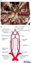
Figure 1. Human angiogram and schematic diagram of cerebral circulation
A, CT scan showing the anatomy of the cerebral arterial supply in humans. The image was kindly provided by Dr Nathan Manghat from University Hospital Bristol Trust. B, labelled schematic representation of A including origins of the circulation from the aortic arch. Figure modified from Schuenke et al. (2010). Abbreviations: A, anterior; R, right; P, posterior; L, left.
Figure 2
Figure 2. Approximate spatial contribution to cerebral blood supply by carotid and vertebral arteries in different species
Schematic representation of the cerebral blood supply contribution by the carotid and vertebral arteries in man (A), rat (B), goat/sheep (C), rabbit (D), dog (E), cat (F) and calf (G). Image based on and adapted from Baldwin & Bell (1963) and Bralet et al. (1977).
Figure 3
Figure 3. Species variation in cerebral artery architecture
Schematic representation of the cerebral arterial tree and difference in contribution to anterior and posterior circulation by the external carotid artery (ECA) and internal carotid artery (ICA) in rats (A), goat/sheep (B) and rabbit (C). Notice the presence of the carotid rete in goat/sheep (B, red arrow), the lack of ICA and the existence of a V‐Oa in the same species. CoW and VA labelled for orientation purposes. Abbreviations: APAr, ascending pharangeal artery residual; BA, basilar artery; CCA, common carotid artery; CoW, Circle of Willis; SA, spinal artery; VA, vertebral artery; V‐Oa, vertebral‐occipital anastomoses. Figure adapted from Daniel et al. (1953), Andersson & Jewell (1956) and Baldwin & Bell (1963).
Figure 4
Figure 4. Parasympathetic innervation to cerebral vasculature
A, schematic overview of the anatomic position of various parasympathetic ganglia that are sources of parasympathetic input to the cerebral arteries. B, the level of parasympathetic innervation to the cerebral arteries varies as shown by VIP innervations in the posterior communicating artery (dense innervation) and basilar artery (moderate innervation); image from Hara et al. (1985); ×172 in original article, reproduced with permission. C, schematic representation of parasympathetic innervations to the rostral and posterior cerebral vasculature. The gradient represents reported density of innervation, cross‐hatched filling indicates unreported innervation. Note that species variation regarding innervation is not depicted in the figure. CmG, carotid mini‐ganglia; CS, cavernous sinus; OG, otic ganglia; PTG; pterygopalatine ganglia; VIP, vasoactive intestinal peptide. Original figure collated from data from Gibbins et al. (1984 a), Hara et al. (1985), Keller et al. (1985), Shimizu (1994), Kadota et al. (1996), Bleys et al. (2001) and Ayajiki et al. (2012).
Figure 5
Figure 5. Electron microscopy image showing the close proximity of sympathetic and parasympathetic terminals on a cerebral artery
Image of the anterior cerebral artery of cat showing the close apposition of a parasympathetic (Ch, cholinergic) and a sympathetic (A, adrenergic) neuron. Schwann cell is visible at the bottom. Image from Edvinsson et al. (1972 b). ×60,000 in original reference, with permission.
Figure 6
Figure 6. Mechanisms of endothelial dependent cross‐talk leading to NO induced vasodilatation
There are multiple sources of nitric oxide production, including the endothelium and nitregic neurones. A, ACh exerts endothelium dependent vasodilatation acting on muscarinic (M) type 3 and/or 5 receptors on endothelial cells inducing an increase in intracellular calcium which leads to NO production via eNOS activity. ACh still evokes a dilatation in denuded vessels suggesting a non‐endothelial source of NO. B, noradrenaline (NA) can stimulate the production of NO from cholinergic/nitrergic neurons via β2 receptors leading to increases in Ca2+ and calcium channel stimulation. The NO diffuses to nearby smooth muscle cells causing relaxation. Modulation of NO release in this system is achieved by ACh‐mediated NO inhibition via prejunctional muscarinic (M) type 2 receptors on nitrergic neurons. Abbreviations: ACh, acetylcholine; cGMP, cyclic guanosine monophosphate; GC, guanylate cyclase; GTP, guanosine triphosphate; NO, nitric oxide; NOS, nitric oxide synthase. Figure based on and adapted from Wahl & Schilling (1993), Zhang et al. (1998 b), Liu et al. (2000) and Si & Lee (2002).

References

    1. Agassandian K, Fazan VS, Margaryan N, Dragon D, Riley J & Talman W (2003). A novel central pathway links arterial baroreceptors and pontine parasympathetic neurons in cerebrovascular control. Cell Mol Neurobiol 23, 463–478. - PMC - PubMed
    1. Akiho H, Chijiiwa Y, Okabe H, Harada N & Nawata H (1995). Interaction between atrial natriuretic peptide and vasoactive intestinal peptide in guinea pig cecal smooth muscle. Gastroenterology 109, 1105–1112. - PubMed
    1. Alpers BJ, Berry RG & Paddison RM (1959). Anatomical studies of the circle of Willis in normal brain. AMA Arch Neurol Psychiatry 81, 409–418. - PubMed
    1. Andersson B & Jewell P (1956). The distribution of carotid and vertebral blood in the brain and spinal cord of the goat. Q J Exp Physiol Cogn Med Sci 41, 462–474.
    1. Ando K (1988). Distribution and origin of vasoactive intestinal polypeptide (VIP)‐immunoreactive, acetylcholinesterase‐positive and adrenergic nerves of the cerebral arteries in the bent‐winged bat (Mammalia: Chiroptera). Cell Tissue Res 251, 345–351. - PubMed

Publication types

MeSH terms

LinkOut - more resources