. 2016 Jun 30:7:12103.
doi: 10.1038/ncomms12103.

Pironetin reacts covalently with cysteine-316 of α-tubulin to destabilize microtubule

Affiliations

Pironetin reacts covalently with cysteine-316 of α-tubulin to destabilize microtubule

Jianhong Yang et al. Nat Commun. .

Abstract

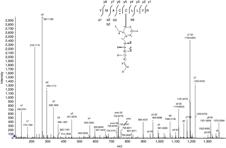
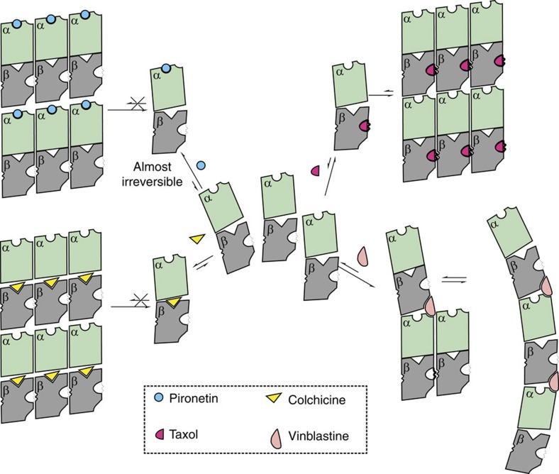
Molecules that alter the normal dynamics of microtubule assembly and disassembly include many anticancer drugs in clinical use. So far all such therapeutics target β-tubulin, and structural biology has explained the basis of their action and permitted design of new drugs. However, by shifting the profile of β-tubulin isoforms, cancer cells become resistant to treatment. Compounds that bind to α-tubulin are less well characterized and unexploited. The natural product pironetin is known to bind to α-tubulin and is a potent inhibitor of microtubule polymerization. Previous reports had identified that pironetin reacts with lysine-352 residue however analogues designed on this model had much lower potency, which was difficult to explain, hindering further development. We report crystallographic and mass spectrometric data that reveal that pironetin forms a covalent bond to cysteine-316 in α-tubulin via a Michael addition reaction. These data provide a basis for the rational design of α-tubulin targeting chemotherapeutics.


Figures

Figure 1
Figure 1. Chemical structure of pironetin and the reaction scheme with cysteine.
Cysteine as a thiol nucleophile undergoes a Michael-type addition to pironetin, in which a protonated base (denoted as B+) or solvent accepts the electron from enolate intermediate.
Figure 2
Figure 2. Structure of the tubulin-RB3-TTL-pironetin complex.
(a) Stereo views of experimental Fo–Fc difference electron density contoured at 3σ in green for pironetin and the side chains of C316 and K352 and the final refined 2Fo–Fc electron density contoured at 1σ in blue. The final refined positions of pironetin, C316 and K352 are shown in stick with carbons coloured cyan for pironetin and green for protein, oxygens red and nitrogens blue. (b) Structure of tubulin-RB3-TTL in complex with pironetin in which pironetin is bound to α2-tubulin. The protein subunits are shown in cartoon. The ligands are shown as spheres with atoms coloured as in a. (c) Close-up view of pironetin in the pocket of α-tubulin monomer. (d) Cross-section view of the surface of the binding pocket of pironetin. K352 previously reported to be modified is shown alongside other residues discussed in the text.
Figure 3
Figure 3. MS/MS fragmentation pattern for pironetin covalently modified peptide from α-tubulin
.The spectrum clearly shows the covalent binding of pironetin to C316 rather than the adjacent C315, which was modified by carbamidomethyl (marked by *). The pironetin containing fragments show characteristic loses of 32 and 50 m/z. Prec denotes precursor ion.
Figure 4
Figure 4. Changes in α-tubulin on pironetin binding.
(a) Comparison of the structures of tubulins in apo form and in complex with pironetin. The tubulin dimers are represented as ribbons. In apo form, α-tubulin is coloured blue and β-tubulin light grey. In the complex with pironetin, α is green and β dark grey. (b) Large main chain changes between apo form (blue) and complex form (green) in H8 and the T7 loop, which links H7 and H8. Two red arrows at L259 and F244 mark the start and stop of the loop. G246, V250 and L252 are shown in sticks and marked with black arrows shift over 8 Å. (c) The catalytic residue E254 of α-tubulin is reoriented along with F255 on pironetin binding. (d) The GTPase activity of tubulin is reduced by pironetin similar to that observed with vinblastine and opposite to the stimulation seen with colchicine.
Figure 5
Figure 5. The molecular mechanism of pironetin and other tubulin-targeting agents to alter the tubulin-microtubule dynamics.
The tubulin-targeting agents colchicine, vinblastine and taxol are shown. This figure is inspired by a previous publication but designed and developed with the data in this study.

References

    1. Alberts B. et al. Molecular Biology of the Cell Garland Science (2008).
    1. Dumontet C. & Jordan M. A. Microtubule-binding agents: a dynamic field of cancer therapeutics. Nat. Rev. Drug Discov. 9, 790–803 (2010). - PMC - PubMed
    1. Kupchan S. M. et al. Maytansine, a novel antileukemic ansa macrolide from Maytenus ovatus. J. Am. Chem. Soc. 94, 1354–1356 (1972). - PubMed
    1. Kavallaris M. Microtubules and resistance to tubulin-binding agents. Nat. Rev. Cancer 10, 194–204 (2010). - PubMed
    1. Nogales E., Wolf S. G., Khan I. A., Luduena R. F. & Downing K. H. Structure of tubulin at 6.5 Å and location of the taxol-binding site. Nature 375, 424–427 (1995). - PubMed

Publication types