. 2016 Aug;24(8):1435-43.
doi: 10.1038/mt.2016.137. Epub 2016 Jun 30.

Adenoviral Delivery of Tumor Necrosis Factor-α and Interleukin-2 Enables Successful Adoptive Cell Therapy of Immunosuppressive Melanoma

Affiliations

Adenoviral Delivery of Tumor Necrosis Factor-α and Interleukin-2 Enables Successful Adoptive Cell Therapy of Immunosuppressive Melanoma

Mikko Siurala et al. Mol Ther. 2016 Aug.

Erratum in

Abstract

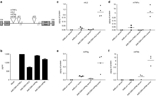
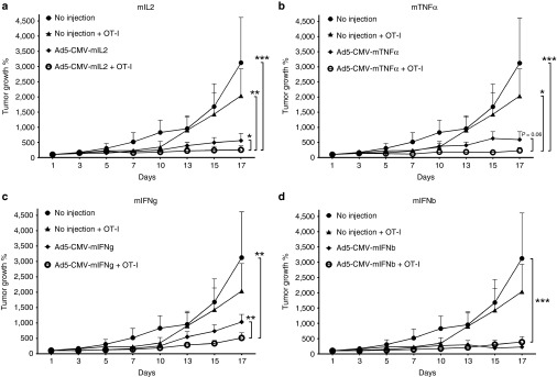
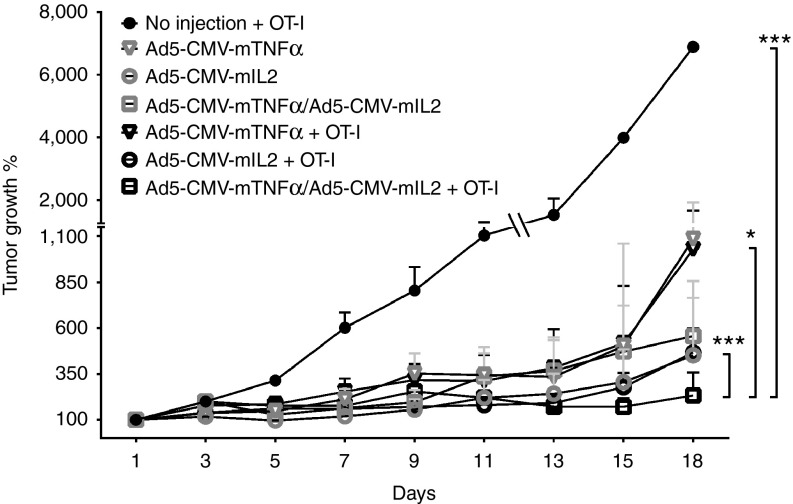
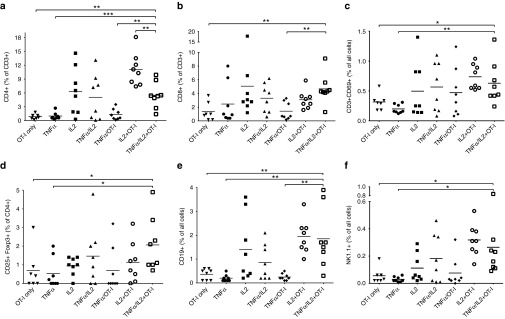
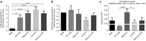
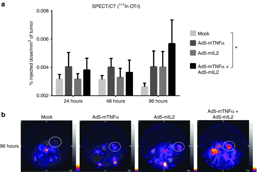
Adoptive T-cell transfer is a promising treatment approach for metastatic cancer, but efficacy in solid tumors has only been achieved with toxic pre- and postconditioning regimens. Thus, adoptive T-cell therapies would benefit from complementary modalities that enable their full potential without excessive toxicity. We aimed to improve the efficacy and safety of adoptive T-cell transfer by using adenoviral vectors for direct delivery of immunomodulatory murine cytokines into B16.OVA melanoma tumors with concomitant T-cell receptor transgenic OT-I T-cell transfer. Armed adenoviruses expressed high local and low systemic levels of cytokine when injected into B16.OVA tumors, suggesting safety of virus-mediated cytokine delivery. Antitumor efficacy was significantly enhanced with adenoviruses coding for murine interleukin-2 (mIL-2) and tumor necrosis factor-α (mTNFα) when compared with T-cell transfer alone or viruses alone. Further improvement in efficacy was achieved with a triple combination of mIL-2, mTNFα, and OT-I T-cells. Mechanistic studies suggest that mIL-2 has an important role in activating T-cells at the tumor, while mTNFα induces chemokine expression. Furthermore, adenovirus treatments enhanced tumor-infiltration of OT-I T-cells as demonstrated by SPECT/CT imaging of (111)In-labeled cells. Our results suggest the utility of cytokine-coding adenoviruses for improving the efficacy of adoptive T-cell therapies.


Figures

Figure 1
Figure 1
Cytokine expression from adenoviral vectors in vitro and in vivo. (a) Schematic of adenovirus construct containing murine cytokine genes under the cytomegalovirus (CMV) promoter. (b) Cytokine concentrations in cell culture supernatants 48 hours after infection. Tumor and serum levels of (c) mIL2, (d) mTNFα, (e) mIFNg, and (f) mIFNb were measured from B16.OVA tumor-bearing mice 72 hours after intratumoral injection of cytokine-coding viruses or unarmed control virus Ad5Luc1. Horizontal lines, mean values.
Figure 2
Figure 2
Antitumor efficacy of cytokine-armed adenoviruses combined with adoptive T-cell transfer. B16-OVA tumor-bearing C57BL/6 mice were administered 1.5 × 106 CD8-enriched OT-I T-cells intraperitoneally on Day 1 with concurrent intratumoral injections of 1 × 109 viral particles of adenoviruses armed with (a) mIL2, (b) mTNFα, (c) mIFNg, and (d) mIFNb. Virus treatments continued every 7 days. Error bars, mean + SEM, n = 5–6. *P < 0.05, **P < 0.01, ***P < 0.001.
Figure 3
Figure 3
Ad5-CMV-mTNFα/Ad5-CMV-mIL2 dual virus combination together with adoptive T-cell transfer. Adenoviruses coding for mTNFα and mIL2 were combined in a 1 to 1 ratio (0.5 × 109 VP of each virus) to treat B16-OVA tumors together with adoptive transfer of 1.5 × 106 CD8-enriched OT-I T-cells. Virus treatments continued every 7 days. Error bars, mean + SEM, n = 8. *P < 0.05, ***P < 0.001.
Figure 4
Figure 4
Tumor-infiltrating lymphocyte subsets following treatment with adenovirus and OT-I combination. Percentages of (a) CD4+ T-cells, (b) CD8+ T-cells, (c) CD3+CD69+ activated T-cells, (d) CD3+CD25+Foxp3+ regulatory T-cells, (e) CD19+ B-cells and (f) NK1.1+ Natural killer cells in B16.OVA tumors treated with 1.5 × 106 CD8-enriched OT-I T-cells and adenoviruses. Horizontal lines, mean values. *P < 0.05, **P < 0.01, ***P < 0.001.
Figure 5
Figure 5
PD1/PD-L1 axis and chemokine expression in B16.OVA tumors. Tumor-bearing mice were killed on Day 7 after treatment with adenoviruses and OT-I T-cells, and the expression of (a) PD-L1 in the tumor and (b) PD1 on CD3+CD8+ TIL were analyzed by flow cytometry. Results are presented as fold change compared to mock treatment. (c) Homogenized tumor lysates were analyzed for the chemokines RANTES, MCP1, MIP-1a, MIP-1b, MIG, and I-TAC. Pooled values of chemokines were normalized to total protein content and presented as picogram per microgram of protein. Error bars, mean + SEM. *P < 0.05, **P < 0.01, ****P < 0.0001.
Figure 6
Figure 6
SPECT/CT imaging of radiolabeled OT-I T-cells adoptively transferred to adenovirus-treated mice. B16.OVA tumor-bearing mice were administered 6 × 106 111Indium-oxine labeled OT-I T-cells intraperitoneally with simultaneous intratumoral injection of cytokine-coding adenoviruses. Mice were imaged on 24, 48, and 96 hours after treatment with a four-headed gamma camera with integrated CT system (nanoSPECT/CT). The results were calculated as percentage of the injected dose per tumor volume (%ID/mm3) (a). Representative SPECT/CT images of 96 hours timepoint with white circles indicating the locations of subcutaneous tumors (b). Error bars, mean + SEM. *P < 0.05, repeated measures analysis of variance.

References

    1. June, CH, Riddell, SR and Schumacher, TN (2015). Adoptive cellular therapy: a race to the finish line. Sci Transl Med 7: 280ps7. - PubMed
    1. Maude, SL, Frey, N, Shaw, PA, Aplenc, R, Barrett, DM, Bunin, NJ et al. (2014). Chimeric antigen receptor T cells for sustained remissions in leukemia. N Engl J Med 371: 1507–1517. - PMC - PubMed
    1. Rosenberg, SA, Yang, JC, Sherry, RM, Kammula, US, Hughes, MS, Phan, GQ et al. (2011). Durable complete responses in heavily pretreated patients with metastatic melanoma using T-cell transfer immunotherapy. Clin Cancer Res 17: 4550–4557. - PMC - PubMed
    1. Johnson, LA, Morgan, RA, Dudley, ME, Cassard, L, Yang, JC, Hughes, MS et al. (2009). Gene therapy with human and mouse T-cell receptors mediates cancer regression and targets normal tissues expressing cognate antigen. Blood 114: 535–546. - PMC - PubMed
    1. Robbins, PF, Morgan, RA, Feldman, SA, Yang, JC, Sherry, RM, Dudley, ME et al. (2011). Tumor regression in patients with metastatic synovial cell sarcoma and melanoma using genetically engineered lymphocytes reactive with NY-ESO-1. J Clin Oncol 29: 917–924. - PMC - PubMed

MeSH terms