. 2016 Aug;14(2):1491-500.
doi: 10.3892/mmr.2016.5415. Epub 2016 Jun 21.

Contribution of human osteoblasts and macrophages to bone matrix degradation and proinflammatory cytokine release after exposure to abrasive endoprosthetic wear particles

Affiliations

Contribution of human osteoblasts and macrophages to bone matrix degradation and proinflammatory cytokine release after exposure to abrasive endoprosthetic wear particles

Anika Jonitz-Heincke et al. Mol Med Rep. 2016 Aug.

Abstract

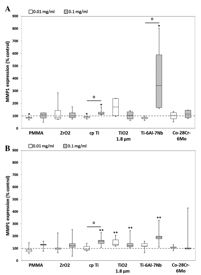
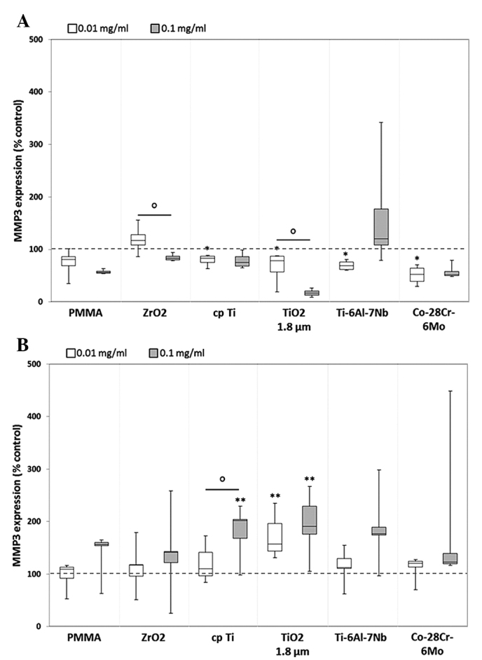
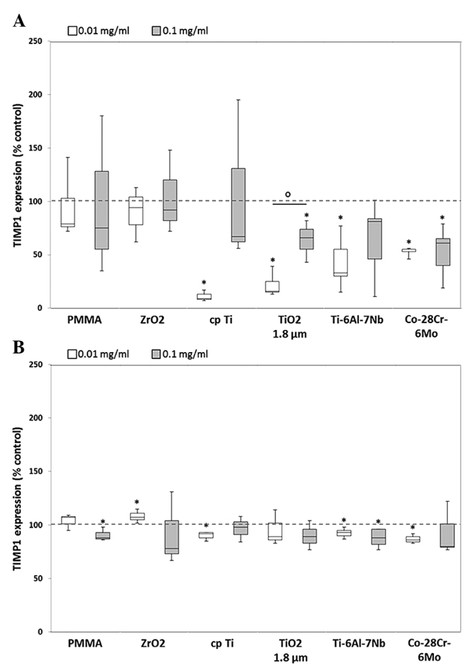
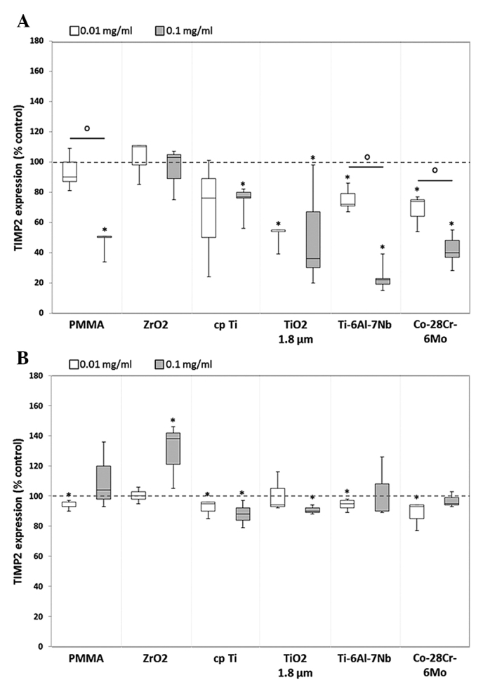
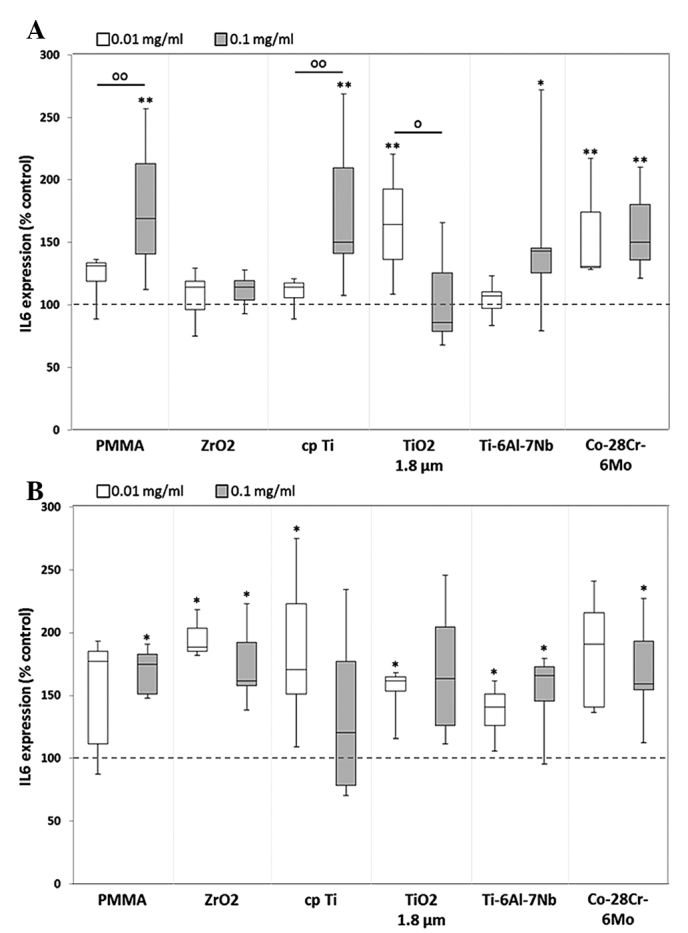
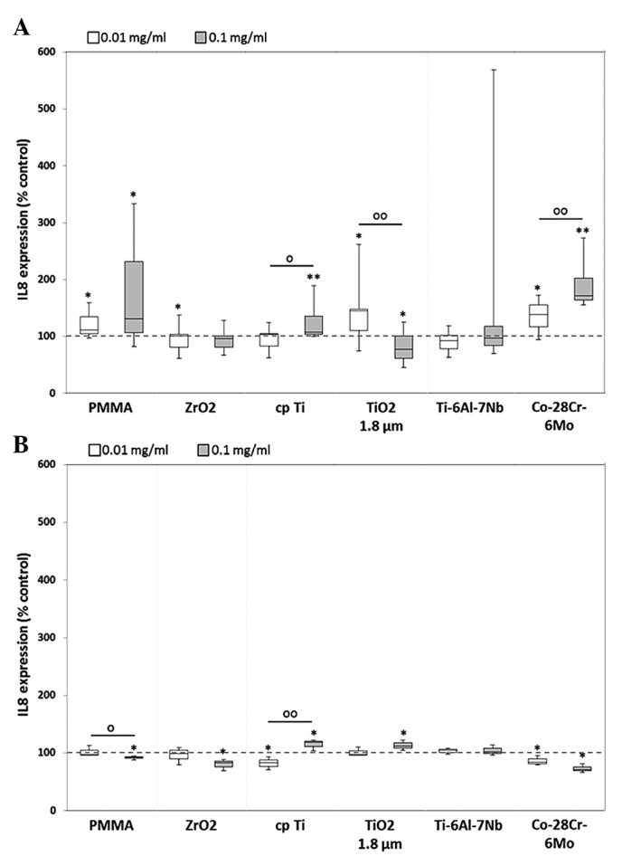
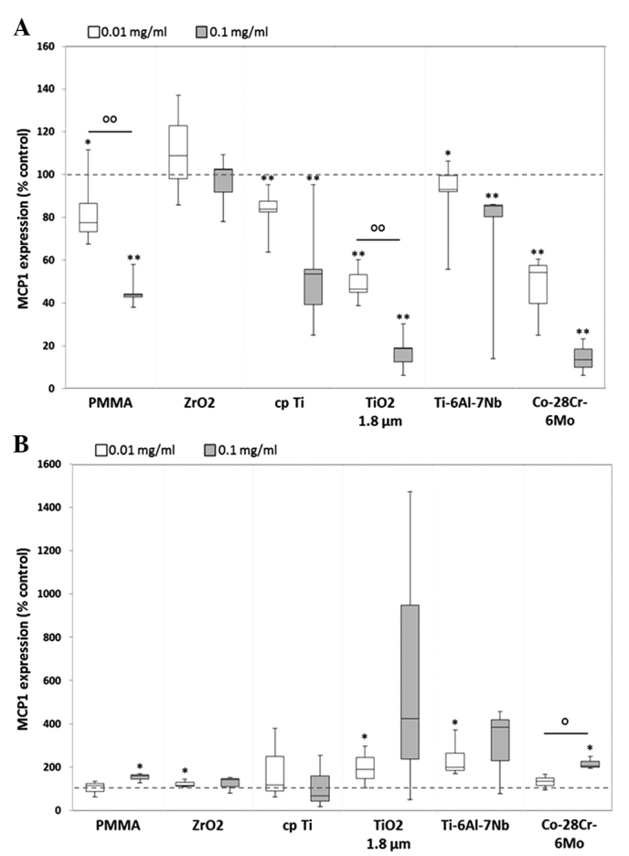
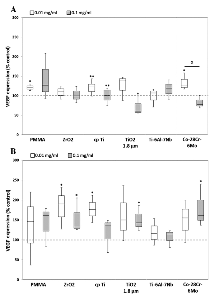
One of the major reasons for failure after total joint arthroplasty is aseptic loosening of the implant. At articulating surfaces, defined as the interface between implant and surrounding bone cement, wear particles can be generated and released into the periprosthetic tissue, resulting in inflammation and osteolysis. The aim of the present study was to evaluate the extent to which osteoblasts and macrophages are responsible for the osteolytic and inflammatory reactions following contact with generated wear particles from Ti‑6Al‑7Nb and Co‑28Cr‑6Mo hip stems. To this end, human osteoblasts and THP‑1 monocytic cells were incubated with the experimentally generated wear particles as well as reference particles (0.01 and 0.1 mg/ml) for 48 h under standard culture conditions. To evaluate the impact of these particles on the two cell types, the release of different bone matrix degrading matrix metalloproteinases (MMPs), tissue inhibitors of MMPs (TIMPs), and relevant cytokines were determined by multiplex enzyme‑linked immunosorbent assays. Following incubation with wear particles, human osteoblasts showed a significant upregulation of MMP1 and MMP8, whereas macrophages reacted with enhanced MMP3, MMP8 and MMP10 production. Moreover, the synthesis of TIMPs 1 and 2 was inhibited. The osteoblasts and macrophages also responded with modified expression of the inflammatory mediators interleukin (IL)‑6, IL‑8, monocyte chemoattractant protein‑1 and vascular endothelial growth factor. These results demonstrate that the release of wear particles affects the release of proinflammatory cytokines and has a negative impact on bone matrix formation during the first 48 h of particle exposure. Human osteoblasts are directly involved in the proinflammatory cascade of bone matrix degradation. The simultaneous activation and recruitment of monocytes/macrophages boosted osteolytic processes in the periprosthetic tissue. By the downregulation of TIMP production and the concomitant upregulation of MMPs as a response to particle exposure, bone formation around implants may be suppressed, resulting in implant failure.


Figures

Figure 1
Figure 1
Release of MMP1 by human (A) osteoblasts and (B) macrophages. Cells were cultivated under standard culture conditions and later treated with the respective particles for 48 h. Subsequently, cell culture supernatants were collected and analyzed using multiplex enzyme-linked immunosorbent assays (osteoblasts, n=4; macrophages, n=3). MMP1 contents are displayed relative to the untreated control. Data are presented as box plots. *P≤0.05 and **P<0.01 vs. particle exposure. °P≤0.05 vs. 0.01 mg/ml (Mann-Whitney U test). MMP, matrix metalloproteinase; PMMA, polymethylmethacrylate; ZrO2, zirconiumoxide; TiO2, titanium dioxide.
Figure 2
Figure 2
Release of MMP3 by human (A) osteoblasts and (B) macrophages. Cells were cultivated under standard culture conditions and later treated with the respective particles for 48 h. Subsequently, cell culture supernatants were collected and analyzed using multiplex enzyme-linked immunosorbent assays (osteoblasts, n=4; macrophages, n=3). MMP3 contents are displayed relative to the untreated control. Data are presented as box plots. *P≤0.05 and **P<0.01 vs. particle exposure. °P≤0.05 vs. 0.01 mg/ml (Mann-Whitney U test). MMP, matrix metalloproteinase; PMMA, polymethylmethacrylate; ZrO2, zirconiumoxide; TiO2, titanium dioxide.
Figure 3
Figure 3
Release of TIMP1 by human (A) osteoblasts and (B) macrophages. Cells were cultivated under standard culture conditions and later treated with the respective particles for 48 h. Subsequently, cell culture supernatants were collected and analyzed using multiplex enzyme-linked immunosorbent assays (osteoblasts, n=4; macrophages, n=3). TIMP1 contents are displayed relative to the untreated control. Data are presented as box plots. *P≤0.05 vs. unstimulated control. °P≤0.05 vs. 0.01 mg/ml (Mann-Whitney U test). TIMP1, tissue inhibitor of matrix metalloproteinase 1; PMMA, polymethyl-methacrylate; ZrO2, zirconiumoxide; TiO2, titanium dioxide.
Figure 4
Figure 4
Release of tissue inhibitor of TIMP2 by human (A) osteoblasts and (B) macrophages. Celles were cultivated under standard culture conditions and later treated with the respective particles for 48 h. Subsequently, cell culture supernatants were collected and analyzed using multiplex enzyme-linked immunosorbent assays (osteoblasts, n=4; macrophages, n=3). TIMP2 contents are displayed relative to the untreated control. Data are presented as box plots. *P≤0.05 vs. particle exposure. °P≤0.05 vs. 0.01 mg/ml (Mann-Whitney U test). TIMP2, tissue inhibitor of matrix metalloproteinase 2; PMMA, polymethylmethacrylate; ZrO2, zirconiumoxide; TiO2, titanium dioxide.
Figure 5
Figure 5
Release of IL-6 by human (A) osteoblasts and (B) macrophages. Cells were cultivated under standard culture conditions and later treated with the respective particles for 48 h. Subsequently, cell supernatants were collected and analyzed using multiplex enzyme-linked immunosorbent assays (osteoblasts, n≥3; macrophages, n=3). IL-6 contents are displayed relative to the untreated control. Data are presented as box plots. *P≤0.05 and **P<0.01 vs. particle exposure. °P≤0.05 and °°P<0.01 vs. 0.01 mg/ml (Mann-Whitney U test). IL-6, interleukkin-6; PMMA, polymethylmethacrylate; ZrO2, zirconiumoxide; TiO2, titanium dioxide.
Figure 6
Figure 6
Release of IL-8 by human (A) osteoblasts and (B) macrophages. Cells were cultivated under standard culture conditions and later treated with the respective particles for 48 h. Subsequently, cell culture supernatants were collected and analyzed using multiplex enzyme-linked immunosorbent assays (osteoblasts, n≥3; macrophages, n=3). IL-8 contents are displayed relative to the untreated control. *P≤0.05 and **P<0.01 vs. particle exposure. °P≤0.05 and °°P<0.01 vs. 0.01 mg/ml (Mann-Whitney U test). IL-8, interleukin 8; PMMA, polymethylmethacrylate; ZrO2, zirconiumoxide; TiO2, titanium dioxide.
Figure 7
Figure 7
Release of MCP1 by human (A) osteoblasts and (B) macrophages. Cells were cultivated under standard culture conditions and later treated with the respective particles for 48 h. Subsequently, cell culture supernatants were collected and analyzed using multiplex enzyme-linked immunosorbent assays (osteoblasts, n ≥3; macrophages, n=3). MCP1 contents are displayed relative to the untreated control. *P≤0.05 and **P<0.01 vs. particle exposure. °P≤0.05 and °°P<0.01 vs. 0.01 mg/ml (Mann-Whitney U test). MCP1, monocyte chemoattractant protein-1; PMMA, polymethylmethacrylate; ZrO2, zirconiumoxide; TiO2, titanium dioxide.
Figure 8
Figure 8
Release of VEGF by human (A) osteoblasts and (B) macrophages. Cells were cultivated under standard culture conditions and later treated with the respective particles for 48 h. Subsequently, cell culture supernatants were collected and analyzed using multiplex enzyme-linked immunosorbent assays (osteoblasts, n ≥3; macrophages, n=3). VEGF contents are displayed relative to the untreated control. *P≤0.05 and **P<0.01 vs. particle exposure. °P≤0.05 vs. 0.01 mg/ml (Mann-Whitney U test). VEGF, vascular endothelial growth factor; PMMA, polymethylmethacrylate; ZrO2, zirconiumoxide; TiO2, titanium dioxide.

References

    1. Bader R, Steinhauser E, Holzwarth U, Schmitt M, Mittelmeier W. A novel test method for evaluation of the abrasive wear behaviour of total hip stems at the interface between implant surface and bone cement. Proc Inst Mech Eng H. 2004;218:223–230. doi: 10.1243/0954411041561009. - DOI - PubMed
    1. Jacobs JJ, Roebuck KA, Archibeck M, Hallab NJ, Glant TT. Osteolysis: Basic science. Clin Orthop Relat Res. 2001;393:71–77. doi: 10.1097/00003086-200112000-00008. - DOI - PubMed
    1. Willert HG, Semlitsch M. Tissue reactions to plastic and metallic wear products of joint endoprostheses. Clin Orthop Relat Res. 1996;333:4–14. doi: 10.1097/00003086-199612000-00002. - DOI - PubMed
    1. Lochner K, Fritsche A, Jonitz A, Hansmann D, Mueller P, Mueller-Hilke B, Bader R. The potential role of human osteoblasts for periprosthetic osteolysis following exposure to wear particles. Int J Mol Med. 2011;28:1055–1063. - PubMed
    1. Syggelos SA, Aletras AJ, Smirlaki I, Skandalis SS. Extracellular matrix degradation and tissue remodeling in periprosthetic loosening and osteolysis: Focus on matrix metalloproteinases, their endogenous tissue inhibitors and the proteasome. Biomed Res Int. 2013;2013:230805. doi: 10.1155/2013/230805. - DOI - PMC - PubMed

MeSH terms