Review
. 2016 Sep;111(4):310-21.
doi: 10.1093/cvr/cvw175. Epub 2016 Jun 29.

Heterogeneity in the lymphatic vascular system and its origin

Affiliations
Review

Heterogeneity in the lymphatic vascular system and its origin

Maria H Ulvmar et al. Cardiovasc Res. 2016 Sep.

Abstract

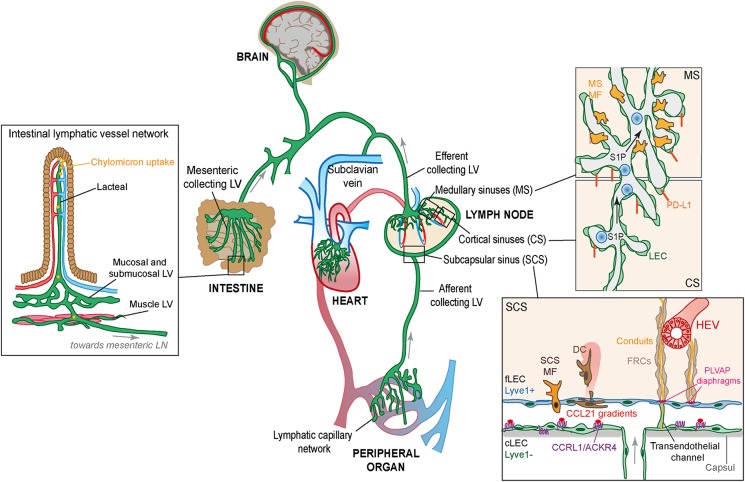
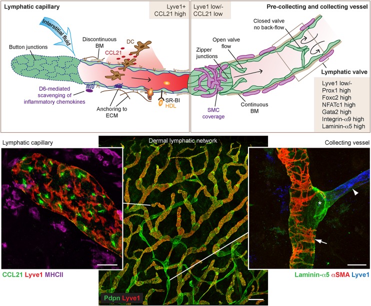
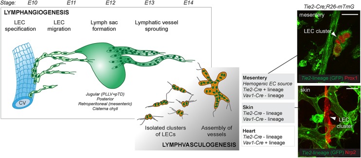
Lymphatic vessels have historically been viewed as passive conduits for fluid and immune cells, but this perspective is increasingly being revised as new functions of lymphatic vessels are revealed. Emerging evidence shows that lymphatic endothelium takes an active part in immune regulation both by antigen presentation and expression of immunomodulatory genes. In addition, lymphatic vessels play an important role in uptake of dietary fat and clearance of cholesterol from peripheral tissues, and they have been implicated in obesity and arteriosclerosis. Lymphatic vessels within different organs and in different physiological and pathological processes show a remarkable plasticity and heterogeneity, reflecting their functional specialization. In addition, lymphatic endothelial cells (LECs) of different organs were recently shown to have alternative developmental origins, which may contribute to the development of the diverse lymphatic vessel and endothelial functions seen in the adult. Here, we discuss recent developments in the understanding of heterogeneity within the lymphatic system considering the organ-specific functional and molecular specialization of LECs and their developmental origin.

Keywords: Haemogenic endothelium; Lymphangiogenesis; Lymphatic vascular development; Lymphatic vessel; Lymphvasculogenesis.


Figures

Figure 1
Figure 1
Organization of the lymphatic vasculature and the specialized lymphatic vessels within the intestine and the lymph node. The lymphatic capillary network (green) absorbs fluid that continuously leaks out from the blood capillary beds and returns it to the blood circulation (blue/red). On the left: The specialized capillary vessels in the intestinal villi called lacteals also take up dietary lipids as chylomicron particles. Both lacteals and capillary vessels in other peripheral organs provide a route for tissue-derived immune cells to the lymph nodes distributed along the collecting vessels; a function that is essential for induction of efficient immune responses and peripheral tolerance. On the right: Molecular and structural features of LECs in different parts of the lymph node (SCS, MS, and CS) are shown. LECs that build the ceiling of the SCSs (cLECs) can be molecularly distinguished from the LECs that form the floor (fLECs) by their low expression of the lymphatic marker Lyve1. cLECs instead express the atypical chemokine receptor CCRL1 (ACKR4) which through scavenging the chemokine CCL21 creates chemokine gradients for migration of tissue-derived DCs (brown) from the SCS into the lymph node parenchyma. Distinct populations of macrophages (MFs; yellow) are associated with SCS and MS but have a common role in scanning the lymph for pathogens and antigens. Small antigens, cytokines, and chemokines can enter the lymph node conduit system that descends from the floor of the SCS in the direction of high endothelial venules (HEVs). The size restriction of the conduit system is determined by PLVAP+ sieve-like diaphragms in transendothelial channels that connect the SCS to the conduits. CS and MS produce S1P essential for immune cell exit from the lymph node and express the immune check point molecule PD-L1 involved in LEC-induced antigen tolerance.
Figure 2
Figure 2
Structural and molecular features of capillary and collecting lymphatic vessels. The blind-ended lymphatic capillaries, designed to remove fluid and soluble molecules from the interstitial space, are characterized by button-like intercellular junctions, discontinuous basement membrane (BM), and anchoring filaments. High capillary expression of the CCR7-ligand CCL21 allows recruitment of DCs. Collecting lymphatic vessels that transport the lymph instead have zipper-like intercellular junctions, continuous basement membrane, smooth muscle cell coverage, and intraluminal valves. The main distinguishing molecular features of the endothelium of capillary vessels, collecting vessels, and valves are depicted in brown boxed areas. Additional functions of the capillary and collecting lymphatic networks include inflammatory chemokine scavenging through expression of the atypical chemokine receptor D6 (ACKR2) and RCT passively or through expression of the cholesterol carrier SR-BI or indirectly through macrophages. Bottom panels show whole-mount immunofluorescence images of mouse ear skin stained with the indicated antibodies. Arrowhead points to a Lyve1 positive capillary and arrowhead to a Lyve1 negative collecting vessel. Valve at a vessel branch point is indicated by an asterisk. Scale bars: 25 µm (capillary and collecting vessels) and 200 µm (lymphatic network).
Figure 3
Figure 3
Key developmental steps in the formation of the embryonic lymphatic vasculature. The first LECs arise through transdifferentiation from venous endothelial cells. Venous-derived LECs exit the veins and form primitive lymphatic structures, so-called lymph sacs, from which vessels sprout further to peripheral organs (lymphangiogenesis). Alongside, in certain organs, non-venous-derived LEC progenitors give rise to clusters of LECs that assemble into vessels (lymphvasculogenesis). Stages (E, embryonic days) of mouse development are shown on the top and key differences in the cellular lineages of non-venous-derived LECs in the mesentery, skin, and heart are depicted. Mesenteric non-venous-derived LECs form from a HemEC source and are traced (GFP+) by the pan-endothelial Tie2-Cre (top panel on the right). In contrast, both the non-venous dermal LECs of lumbar skin (bottom panel on the right) and part of the cardiac lymphatic vasculature form from Tie2 negative lineage (GFP-), and their precise cellular origin within the embryo still remains to be determined. Scale bars: 25 µm.

References

    1. Card CM, Yu SS, Swartz MA. Emerging roles of lymphatic endothelium in regulating adaptive immunity. J Clin Invest 2014;124:943–952. - PMC - PubMed
    1. Aebischer D, Iolyeva M, Halin C. The inflammatory response of lymphatic endothelium. Angiogenesis 2014;17:383–393. - PubMed
    1. Randolph GJ, Miller NE. Lymphatic transport of high-density lipoproteins and chylomicrons. J Clin Invest 2014;124:929–935. - PMC - PubMed
    1. Dixon JB. Lymphatic lipid transport: sewer or subway? Trends Endocrinol Metab 2010;21:480–487. - PMC - PubMed
    1. Aspelund A, Robciuc MR, Karaman S, Makinen T, Alitalo K. Lymphatic system in cardiovascular medicine. Circ Res 2016;118:515–530. - PubMed

Publication types

MeSH terms

Substances