. 2016 Jul 12;113(28):7786-91.
doi: 10.1073/pnas.1608061113. Epub 2016 Jun 28.

Gpr161 anchoring of PKA consolidates GPCR and cAMP signaling

Affiliations

Gpr161 anchoring of PKA consolidates GPCR and cAMP signaling

Verena A Bachmann et al. Proc Natl Acad Sci U S A. .

Abstract

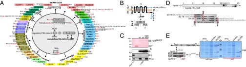
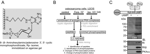
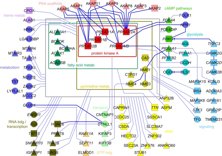
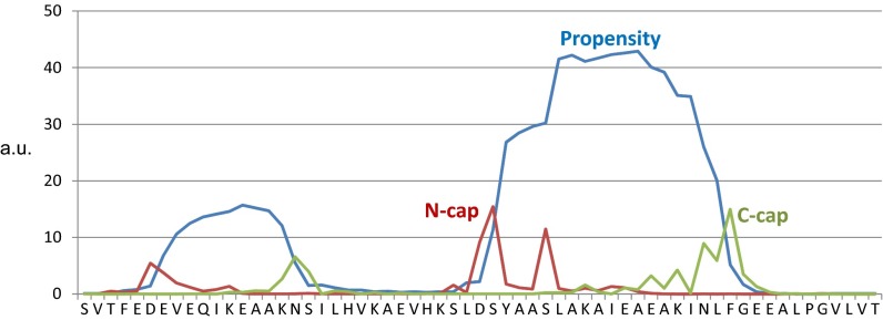
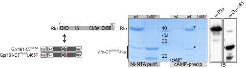
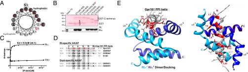
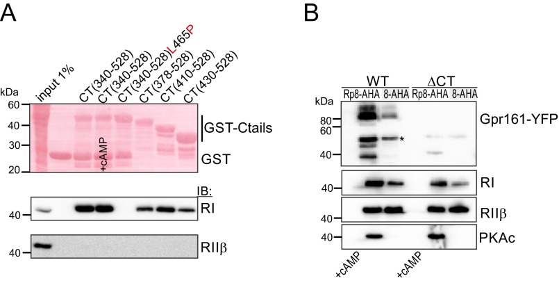
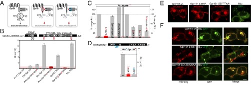
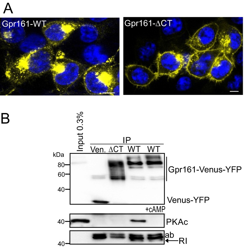
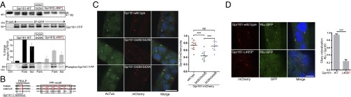
Scaffolding proteins organize the information flow from activated G protein-coupled receptors (GPCRs) to intracellular effector cascades both spatially and temporally. By this means, signaling scaffolds, such as A-kinase anchoring proteins (AKAPs), compartmentalize kinase activity and ensure substrate selectivity. Using a phosphoproteomics approach we identified a physical and functional connection between protein kinase A (PKA) and Gpr161 (an orphan GPCR) signaling. We show that Gpr161 functions as a selective high-affinity AKAP for type I PKA regulatory subunits (RI). Using cell-based reporters to map protein-protein interactions, we discovered that RI binds directly and selectively to a hydrophobic protein-protein interaction interface in the cytoplasmic carboxyl-terminal tail of Gpr161. Furthermore, our data demonstrate that a binary complex between Gpr161 and RI promotes the compartmentalization of Gpr161 to the plasma membrane. Moreover, we show that Gpr161, functioning as an AKAP, recruits PKA RI to primary cilia in zebrafish embryos. We also show that Gpr161 is a target of PKA phosphorylation, and that mutation of the PKA phosphorylation site affects ciliary receptor localization. Thus, we propose that Gpr161 is itself an AKAP and that the cAMP-sensing Gpr161:PKA complex acts as cilium-compartmentalized signalosome, a concept that now needs to be considered in the analyzing, interpreting, and pharmaceutical targeting of PKA-associated functions.

Keywords: interaction network; molecular interactions; phosphorylation; primary cilium; scaffolding function.


Conflict of interest statement

The authors declare no conflict of interest.

Figures

Fig. 1.
Fig. 1.
PKA phosphoprotein interactome and Gpr161:RI interaction. (A) Phosphoproteins of affinity-purified PKA complexes are listed. Underlining marks previously described p-sites; red labeling highlights PKA group sites with a phosphorylated RxxS/T sequence motif. (B) Schematic illustration of Gpr161 trafficking and PKA signaling; cAMP binds R subunits and activates PKAc. (C) GST pulldown experiments of endogenous PKA subunits from HEK293 cell lysates in the presence or absence of 5 mM cAMP using GST and GST-CT (murine Gpr161-Carboxy-terminus340–528) hybrid proteins. (D) Spotted peptides (25-mers, 15-aa overlap) of human Gpr161-CT were overlaid with recombinant RI. Immunoblotting (IB) has been performed with a monoclonal anti-RI antibody. The helix propensity of this part of Gpr161-CT (flanked by L458 and L477) is shown. Indication of L465. (E) Following bacterial coexpression of his6-tagged CT378–528 (wild-type and L465P) and untagged RIα we purified complexes using Ni-NTA resin. Representative experiment from n = 3. Asterisks (*) indicate specific Coomassie brilliant blue-stained bands.
Fig. S1.
Fig. S1.
Strategy to isolate endogenous PKA complexes using a PKA specific cAMP-agarose resin. (A) Structure of Rp-8AHA-cAMP agarose. (B) Flowchart of the proteomics and phosphoproteomics approach. Affinity-isolated PKA complexes from U2OS cells were analyzed using LC-MS/MS followed by semiquantitative interrelationship analyses. (C) Selective enrichment of endogenous PKA subunits from the osteosarcoma cell line U2OS. As negative control, we added excess of cAMP to mask cAMP-binding sites in R subunits for precipitation.
Fig. S2.
Fig. S2.
PPI network emanating from PKA. Following phosphoproteomic analyses using LC-MS/MS, we generated a static PPI network of the confident set of PKA interacting proteins. We neglected applied pharmacological applications for the construction of the macromolecular PKA PPI network. Dark lines indicate known interactions. Triangles indicate identified phosphopeptides; circles indicate identified peptides; squares indicate identified peptides and phosphopeptides from the same protein or protein isoform. With italic lettering we indicate proteins with nucleotide binding capacity, including ATP, cAMP, cGMP, FAD, NAD, and GTP binding.
Fig. S3.
Fig. S3.
Helix propensity prediction. Helix propensity predictions of human Gpr161 were measured with Agadir, an algorithm to predict the helical content of peptides (54).
Fig. S4.
Fig. S4.
Bacterial expression and interaction analyzes of Gpr161-CT and RIα. Following bacterial coexpression of hexa-histidin (his6) and s-tag tagged CT378–528 (wild-type and L465P) and untagged RIα (schematics are shown), we purified complexes using Ni-NTA resin. Subsequent to imidazol elutions, CT378–528-bound complexes have been subjected to cAMP precipitations (±excess of cAMP; 5 mM). Asterisks (*) indicate specific Coomassie brilliant blue-stained bands. IB (with GPR161 and RI antibodies) has been performed to identify protein bands.
Fig. 2.
Fig. 2.
Interaction of Gpr161:RIα in vitro and in silico. (A) Helical wheel projection of the helix propensity of Gpr161-CT from L458 to L477 (murine Gpr161 numbering; helix numbering 1–20). Red labeling indicates conserved amino acids potentially involved in PPI with RI. (B) GST pulldown experiments of recombinant RIα in the presence or absence of 5 mM cAMP using indicated GST-CT fusions of Gpr161 including the CT340–528-L465P mutant. (C) Binding analyzes of full-length RIα (●) or full-length RIIβ (■) to the 5-carboxyfluorescein (5-FAM)–labeled peptide of Gpr161 (amino acids 457–481; 25-mer) by steady-state fluorescence anisotropy measurements. ±SD from n = 3 independent experiments. (D) Structure-based alignment of Gpr161 (murine) with RI-specific and dual-specific AKAPs (human). The four binding pockets are highlighted in gray. Conserved and possibly PPI-involved amino acids are shown in bold. Gpr161 residues that seem to participate in the interaction with the hydrophobic groove of the RI-D/D domain are highlighted in red. Identity (*) and similarities (: and .) are indicated. (E) Structural model of RIα D/D domain in complex with Gpr161-CT456-478 peptide. Crystal structure of RIα D/D-domain:DAKAP-2 peptide (PDB ID code 3IM4) was used to build the model. The DAKAP-2 sequence was substituted with the predicted amphipathic helix sequence of Gpr161-CT456–478. Side and top view of the model are shown. The monomers of the RIα D/D domain are depicted in blue and the Gpr161 helix is shown gray with PPI-relevant amino acids highlighted in red (helix numbering 1–20).
Fig. S5.
Fig. S5.
Biochemical interaction studies of Gpr161 and PKA. (A) GST pulldown analyzes of endogenous PKA subunits from HEK293 cell lysates in the presence or absence of 5 mM cAMP using GST and GST-CT variants. (B) cAMP-precipitation of endogenous PKA complexes and overexpressed Gpr161 variants tagged with Venus-YFP using two types of cAMP agarose. We have enriched PKA holoenzyme complexes using Rp-8-AHA-cAMP and activated PKA regulatory complexes using 8-AHA-cAMP. In the negative control experiment we added an excess of cAMP (5 mM) to the lysates to mask the cAMP binding sites in the R subunits for precipitation. An asterisk (*) indicates a degradation product of Gpr161-YFP.
Fig. S6.
Fig. S6.
AKAP-peptide:RIα interactions. Spotted peptide sequences directly derived from amphipathic helices of indicated AKAPs or in silico-designed peptides (29) were overlaid with recombinant RIα. We have integrated proline mutations to control RIα binding. IB has been performed with a monoclonal anti-RI antibody.
Fig. 3.
Fig. 3.
Cellular PPIs and localization of Gpr161 variants. (A) Schematic illustration of the Rluc-PCA biosensor strategy to quantify PPIs of wild-type and mutated Gpr161 and RI in vivo. Mutated domains are highlighted in red/blue. (B) Shown are conserved sequence elements in the Gpr161-CT. Impact of L465P mutation of Gpr161-F[1]/[2] on complex formation with RIα-F[1]/[2] (±SEM; representative of n = 3 independent experiments; murine Gpr1611–528; NP_001297359.1). (C) Impact of Gpr161 mutations of the flanking Leu of the PPI-motif and the PKA phosphorylation consensus site on RIα:Gpr161 PPI. Read out: Rluc PCA (±SEM of at least n = 4 independent experiments). (D) Impact of the L50R mutation on RIβ:Gpr161 PPI; Rluc PCA measurements (±SEM; representative of n = 3). (E) Subcellular localization of mCherry-tagged Gpr161 or GFP-tagged RIα hybrid proteins in HEK293 cells. (Scale bar, 5 μm.) (F) Subcellular localization of coexpressed mCherry-tagged Gpr161 variants and GFP-tagged RIα in HEK293 cells. (Scale bar, 5 μm.)
Fig. S7.
Fig. S7.
IP and localization of Gpr161. (A) Subcellular localization of Venus-YFP tagged Gpr161 fusion proteins in HEK293 cells. (Scale bars, 5 μm.) (B) IP of Venus-YFP tagged Gpr161 variants expressed in HEK293 cells (representative of n = 3 independent experiments).
Fig. 4.
Fig. 4.
Phosphorylation and ciliary localization of Gpr161:RIα complexes. (A) IP of Venus-YFP–tagged Gpr161 variants expressed in HEK293 cells following treatments with Forskolin (20 µM, 10 min) and isoproterenol (1 µM, 10 min). Densitometric quantification of n = 4 independent experiments, ±SEM; phospho-(K/R)(K/R)X(S*/T*) specific antibody. The IB with the RI antibody is taken from a different experiment (better separation of antibody and RI). (B) Sequence comparison of AKAP and phosphorylation motifs from human and zebrafish Gpr161-CT. (C) Subcellular localization of indicated Gpr161-mCherry variants and acetylated-Tubulin in zebrafish. (Scale bar, 10 µm.) The graph shows the ratio of Gpr161-positive cilia and the total number of cilia in a minimum of three sections from three independent experiments. P values were calculated using one-way ANOVA and Tukey's multiple-comparison post hoc test (***P < 0.001). Shown are the individual ratios (±SD) of Gpr161-mCherry wild-type (12 sections, 578 cilia), Gpr161-mCherry S428D/S429D (9 sections, 584 cilia), and Gpr161-mCherry S428A/S429A (9 sections, 415 cilia). (D) Coexpression of Gpr161-mCherry and RIα-GFP in zebrafish. The graph shows the ratio of RIα, Gpr161 double-positive cilia over the total number of Gpr161-mCherry-positive cilia (mean ± SEM, Gpr161-mCherry wild-type n = 7 embryos, Gpr161-mCherry L465P n = 6 embryos). ***P < 0.001 using two-tailed unpaired Student’s t test. (Scale bar, 5 µm.)

References

    1. Langeberg LK, Scott JD. Signalling scaffolds and local organization of cellular behaviour. Nat Rev Mol Cell Biol. 2015;16(4):232–244. - PMC - PubMed
    1. Shukla AK, Xiao K, Lefkowitz RJ. Emerging paradigms of β-arrestin-dependent seven transmembrane receptor signaling. Trends Biochem Sci. 2011;36(9):457–469. - PMC - PubMed
    1. Good MC, Zalatan JG, Lim WA. Scaffold proteins: Hubs for controlling the flow of cellular information. Science. 2011;332(6030):680–686. - PMC - PubMed
    1. Ritter SL, Hall RA. Fine-tuning of GPCR activity by receptor-interacting proteins. Nat Rev Mol Cell Biol. 2009;10(12):819–830. - PMC - PubMed
    1. Scott JD, Dessauer CW, Taskén K. Creating order from chaos: Cellular regulation by kinase anchoring. Annu Rev Pharmacol Toxicol. 2013;53:187–210. - PMC - PubMed

Publication types