Review
. 2016 Jun 30;20(1):150.
doi: 10.1186/s13054-016-1329-y.

"Awake" extracorporeal membrane oxygenation (ECMO): pathophysiology, technical considerations, and clinical pioneering

Affiliations
Review

"Awake" extracorporeal membrane oxygenation (ECMO): pathophysiology, technical considerations, and clinical pioneering

Thomas Langer et al. Crit Care. .

Abstract

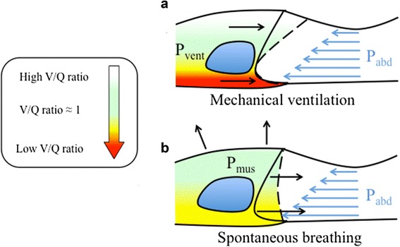
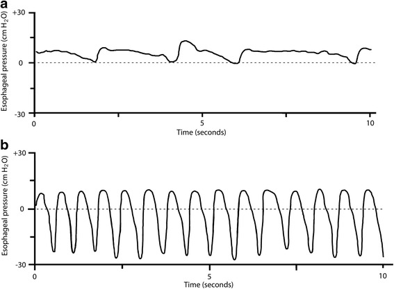
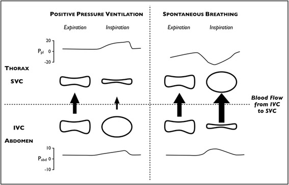
Venovenous extracorporeal membrane oxygenation (vv-ECMO) has been classically employed as a rescue therapy for patients with respiratory failure not treatable with conventional mechanical ventilation alone. In recent years, however, the timing of ECMO initiation has been readdressed and ECMO is often started earlier in the time course of respiratory failure. Furthermore, some centers are starting to use ECMO as a first line of treatment, i.e., as an alternative to invasive mechanical ventilation in awake, non-intubated, spontaneously breathing patients with respiratory failure ("awake" ECMO). There is a strong rationale for this type of respiratory support as it avoids several side effects related to sedation, intubation, and mechanical ventilation. However, the complexity of the patient-ECMO interactions, the difficulties related to respiratory monitoring, and the management of an awake patient on extracorporeal support together pose a major challenge for the intensive care unit staff. Here, we review the use of vv-ECMO in awake, spontaneously breathing patients with respiratory failure, highlighting the pros and cons of this approach, analyzing the pathophysiology of patient-ECMO interactions, detailing some of the technical aspects, and summarizing the initial clinical experience gained over the past years.


Figures

Fig. 1
Fig. 1
Diaphragm motion and ventilation/perfusion distribution in the awake and in the anesthetized subject. The lung ventilation-to-perfusion ratio (V/Q) is color-coded from white (high V/Q), to green (V/Q ≈ 1), to red (low V/Q). Diaphragm shape at end expiration (continuous line) and end inspiration (dashed line) in the supine position is shown. Intra-abdominal pressure increases in the ventro-dorsal direction due to gravity (blue arrows) and displaces the dorsal part of the diaphragm more cephalad than the ventral part at end expiration. During mechanical ventilation the pressure applied by the mechanical ventilator displaces the ventral part of the diaphragm, which faces less intra-abdominal pressure, more than the dorsal part (passive movement). Ventilation will thus be distributed preferentially to the ventral lung regions, increasing the ventilation-to-perfusion ratio (V/Q) of these areas. In contrast, dorsal lung regions will receive less ventilation and their V/Q will be lower (a). During spontaneous breathing (either assisted or unassisted), both the ventral and the dorsal part of the diaphragm move (active contraction). Ventilation will distribute more homogeneously along the ventro-dorsal axis of the lung and will more closely match perfusion (V/Q ≈ 1) (b)
Fig. 2
Fig. 2
Esophageal pressure swings during spontaneous breathing in normal conditions and with ARDS. a Esophageal pressure (Pes) trace of an awake, spontaneously breathing sheep with healthy lungs. Esophageal pressure swings (∆Pes) are around 4–6 cmH2O and the respiratory rate is around 14–18 breaths per minute. b Pes trace of an awake, spontaneously breathing sheep with oleic acid-induced ARDS. Measured ∆Pes values are around 20–30 cmH2O and respiratory rate is greatly increased. Personal experimental data of Thomas Langer and Andriy Batchinsky [49]
Fig. 3
Fig. 3
Shape of the intra- and extrathoracic veins in different transmural pressure conditions (heart–lung interactions). Changes in pleural pressure (P pl), abdominal pressure (P abd), the shape of the superior and inferior venae cavae (SVC and IVC, respectively), and the amount of blood flow (arrows) from the abdomen to the thorax during mechanical ventilation (left panel) and spontaneous breathing (right panel). During positive pressure ventilation, the increased pleural pressure squeezes the SVC and reduces blood flow from the abdominal compartment. This induces a distention of the IVC, favoring blood drainage to the extracorporeal circuit. During spontaneous breathing with high inspiratory effort, the significant decrease in pleural pressure dilates the SVC and increases blood flow from the abdominal compartment. This may induce a collapse of the IVC, hindering blood drainage to the extracorporeal circuit

References

    1. Gattinoni L, Pesenti A, Mascheroni D, Marcolin R, Fumagalli R, Rossi F, et al. Low-frequency positive-pressure ventilation with extracorporeal CO2 removal in severe acute respiratory failure. JAMA. 1986;256:881–6. doi: 10.1001/jama.1986.03380070087025. - DOI - PubMed
    1. Morris AH, Wallace CJ, Menlove RL, Clemmer TP, Orme JF, Jr, Weaver LK, et al. Randomized clinical trial of pressure-controlled inverse ratio ventilation and extracorporeal CO2 removal for adult respiratory distress syndrome. Am J Respir Crit Care Med. 1994;149:295–305. doi: 10.1164/ajrccm.149.2.8306022. - DOI - PubMed
    1. Davies A, Jones D, Bailey M, Beca J, Bellomo R, Blackwell N, et al. Extracorporeal membrane oxygenation for 2009 influenza A (H1N1) acute respiratory distress syndrome. JAMA. 2009;2009(302):1888–95. - PubMed
    1. Gattinoni L, Carlesso E, Langer T. Extracorporeal membrane oxygenation. Crit Care. 2011;15:243. doi: 10.1186/cc10490. - DOI - PMC - PubMed
    1. Peek GJ, Mugford M, Tiruvoipati R, Wilson A, Allen E, Thalanany MM, et al. Efficacy and economic assessment of conventional ventilatory support versus extracorporeal membrane oxygenation for severe adult respiratory failure (CESAR): a multicentre randomised controlled trial. Lancet. 2009;374:1351–63. doi: 10.1016/S0140-6736(09)61069-2. - DOI - PubMed

Publication types

MeSH terms