Review
. 2016 Aug 18;128(7):893-901.
doi: 10.1182/blood-2016-04-636456. Epub 2016 Jun 29.

Vascular thiol isomerases

Affiliations
Review

Vascular thiol isomerases

Robert Flaumenhaft et al. Blood. .

Abstract

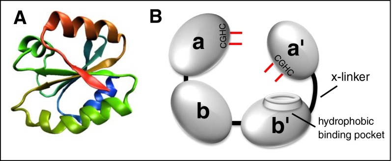
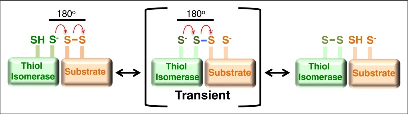
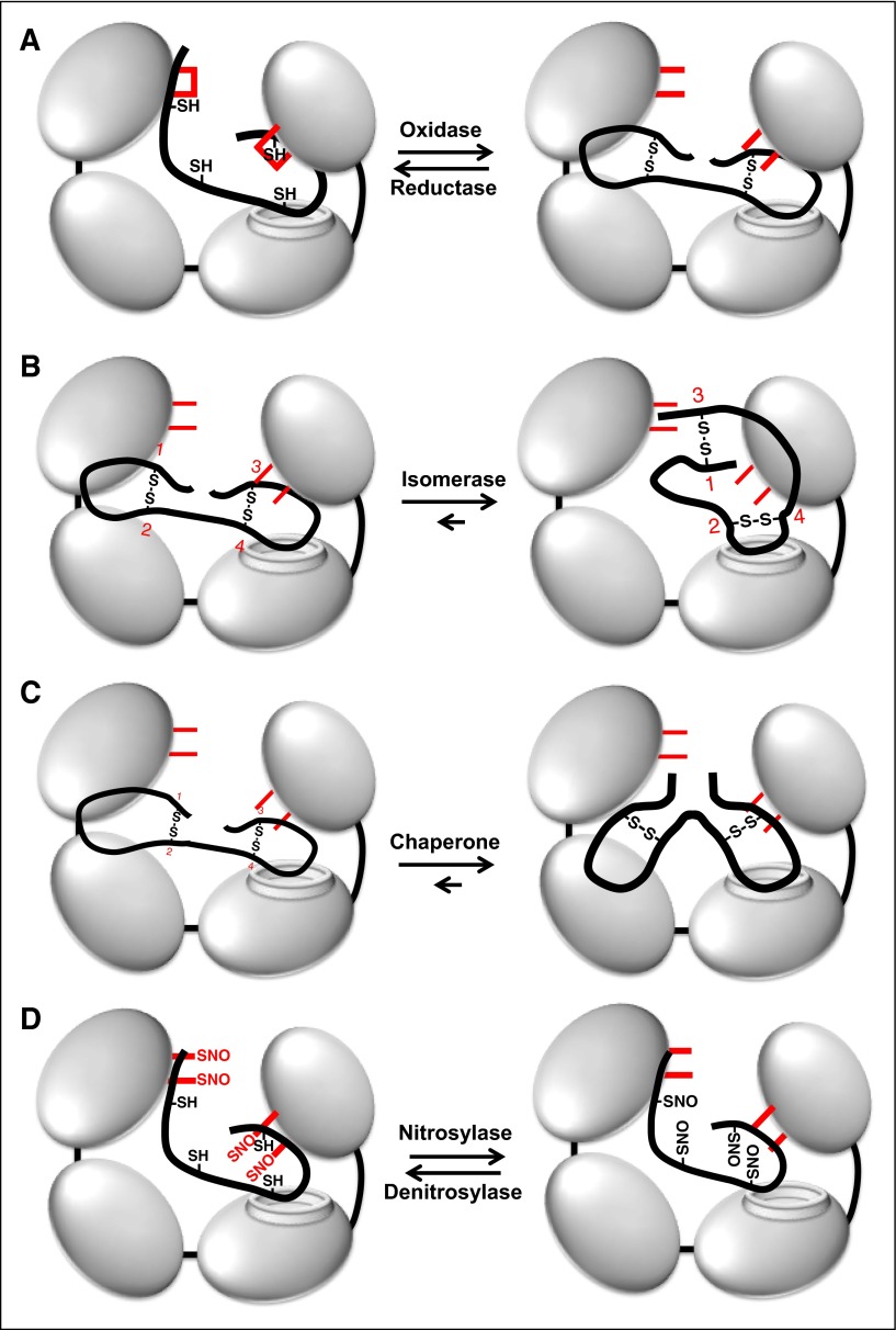
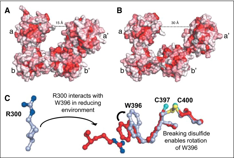
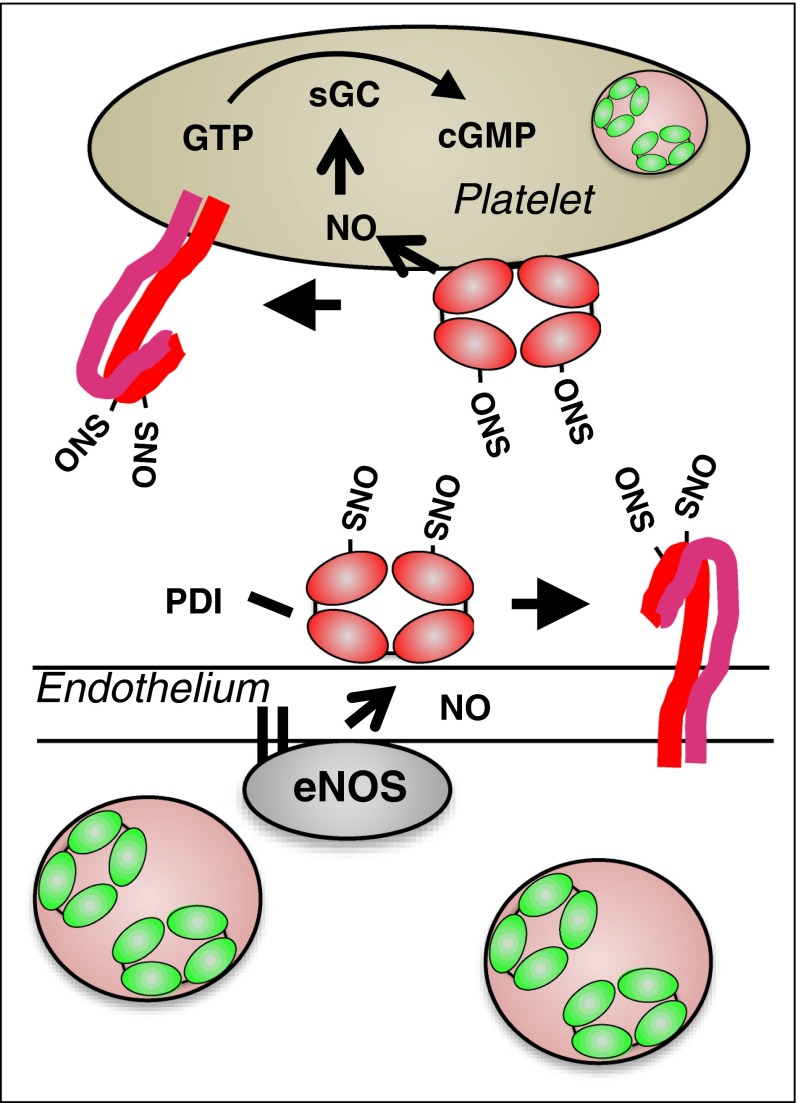
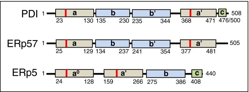
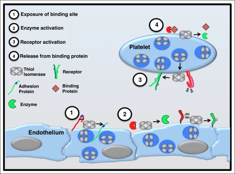
Thiol isomerases are multifunctional enzymes that influence protein structure via their oxidoreductase, isomerase, and chaperone activities. These enzymes localize at high concentrations in the endoplasmic reticulum of all eukaryotic cells where they serve an essential function in folding nascent proteins. However, thiol isomerases can escape endoplasmic retention and be secreted and localized on plasma membranes. Several thiol isomerases including protein disulfide isomerase, ERp57, and ERp5 are secreted by and localize to the membranes of platelets and endothelial cells. These vascular thiol isomerases are released following vessel injury and participate in thrombus formation. Although most of the activities of vascular thiol isomerases that contribute to thrombus formation are yet to be defined at the molecular level, allosteric disulfide bonds that are modified by thiol isomerases have been described in substrates such as αIIbβ3, αvβ3, GPIbα, tissue factor, and thrombospondin. Vascular thiol isomerases also act as redox sensors. They respond to the local redox environment and influence S-nitrosylation of surface proteins on platelets and endothelial cells. Despite our rudimentary understanding of the mechanisms by which thiol isomerases control vascular function, the clinical utility of targeting them in thrombotic disorders is already being explored in clinical trials.


Figures

Figure 1
Figure 1
The architecture of thiol isomerases. Thiol isomerases consist of tandem domains containing a thioredoxin-fold. (A) The structure of the thioredoxin-fold. (B) PDI is formed from 4 tandem domains containing thioredoxin-like folds that assume a U-shape. The 2 catalytic domains (a and a′) contain the CGHC motifs that contain the catalytic cysteines (shown in red). The catalytic cysteines face one another and are responsible for oxidoreductive activities. The substrate binding domains (b and b′) form the bottom of the U-shape. The b′ domain contains a hydrophobic binding pocket that is primarily responsible for substrate binding. The b′ and a′ domains are connected by a flexible 19-amino-acid peptide termed the x-linker.
Figure 2
Figure 2
Mechanism of oxidoreductive cleavage by thiol isomerases. The reduced thiol isomerase aligns with a substrate that contains a disulfide bond such that the free thiolate forms an 180° angle with the vector of the disulfide. A second-order nucleophilic substitution SN2-type reaction then occurs in which the active site sulfur ion nucleophile of the thiol isomerase attacks the adjacent sulfur atoms of the disulfide bond. This nucleophilic substitution results in a transient mixed disulfide (blue). The mixed disulfide spontaneously decomposes, resulting in the formation of a disulfide bond in the thiol isomerase and reduction of the disulfide bond in the substrate.
Figure 3
Figure 3
Multiple functions of PDI. (A) PDI is an oxidoreductase that can both form (oxidase) and cleave (reductase) disulfide bonds. Active site cysteines are shown in red. Note that the catalytic cysteines are either reduced or oxidized during these processes. (B) Through its oxidoreductase activity, PDI acts as an isomerase changing disulfide bond patterns within a protein. In this example, PDI modifies disulfides formed between cysteines 1-2 and 3-4 into disulfides formed between 1-3 and 2-4 (numbering in red). Note that there is no net change in the redox state of the catalytic cysteines on PDI. (C) PDI can act as a chaperone, modifying the conformation of a protein independent of its oxidoreductase and chaperone activities. The redox state of the catalytic cysteines is unchanged. (D) The active site cysteines within PDI can also become S-nitrosylated. PDI can then transfer NO to substrates, thus acting as a nitrosylase. PDI can also act as a denitrosylase, removing NO from substrates. S-nitrosylation typically inactivates substrates, whereas denitrosylation activates substrates.
Figure 4
Figure 4
Redox sensitivity of PDI. (A) In a reducing environment, PDI assumes a compact structure characterized by a 15-Å distance between the a and a′ domains. (B) In an oxidizing environment, the distance between the a and a′ domain increases to 30 Å. The increase in distance results from loss of interactions between the b′ and a′ domains that allows movement of the x-linker and a′ domains. Adapted from Wang et al. (C) In a reducing environment, the disulfide bond between C397 and C400 is broken, enabling the rotation of W396. This rotation positions W396 so that it can interact with R300. The interaction between W396 and R300 brings the a′ domain in closer proximity to the b′ domain, facilitating additional interactions that lead to a more compact structure. Adapted from Wang et al.
Figure 5
Figure 5
Proposed model for the role of PDI in regulating vascular NO activity. In the quiescent vasculature, when NO levels are high, PDI may be S-nitrosylated. Endothelial SNO-PDI could transfer NO to surface proteins, thereby inhibiting their activity, or transfer NO intracellularly. Intracellular NO could bind to guanylyl cyclase, stimulating the generation of cGMP and contributing the platelet quiescence.
Figure 6
Figure 6
Domain structures of vascular thiol isomerases. (A) The domain structures of thiol isomerases with known activity in thrombus formation (PDI, ERp57, and ERp5) are shown. Catalytic domains (tan), binding domains (blue), and c domains (green) are indicated. The location of the CGHC motif is indicated in red. Numbers indicate the amino acid position in the mature protein.
Figure 7
Figure 7
Functional consequences of thiol isomerase-mediated disulfide bond modifications. Thiol isomerases are secreted from platelets and endothelium following cell activation and mediate modifications of functional disulfide bonds in multiple substrates. Several categories of modifications are observed. (1) Exposure of binding sites in adhesion proteins. Cleavage of disulfide bonds can reveal cryptic binding sites such as RGD sequences (shown in blue) enabling a nonbinding adhesion protein (red) to adhere to binding partners on the cell surface (green). Thrombospondin is an adhesion protein that is modified in this manner. (2) Activation of enzyme function. Functional disulfide bond cleavage could also expose an encrypted active site within an enzyme or modify its conformation, converting the enzyme to a more active conformation. Conversely, formation of a disulfide bond could activate an enzyme or coenzyme (eg, tissue factor).,, (3) Receptor activation. Disulfide bond shuffling could contribute to the formation or stabilization of the active conformation of cell surface receptors (eg, αIIbβ3)., (4) Release from binding protein. Cleavage of a disulfide bond could release a protein from its binding partner, thereby activating the protein (eg, TGF-β).

References

    1. Sela M, White FH, Jr, Anfinsen CB. Reductive cleavage of disulfide bridges in ribonuclease. Science. 1957;125(3250):691–692. - PubMed
    1. Dani HM, Fielder JA, Rabin BR. Degranulation and sex-specific regranulation of reticular membranes from rat liver as studied using a spectrophotometric assay of protein disulphide isomerase. FEBS Lett. 1976;65(3):377–382. - PubMed
    1. Goldberger RF, Epstein CJ, Anfinsen CB. Acceleration of reactivation of reduced bovine pancreatic ribonuclease by a microsomal system from rat liver. J Biol Chem. 1963;238:628–635. - PubMed
    1. Bulleid NJ, Freedman RB. Defective co-translational formation of disulphide bonds in protein disulphide-isomerase-deficient microsomes. Nature. 1988;335(6191):649–651. - PubMed
    1. Ellgaard L, Ruddock LW. The human protein disulphide isomerase family: substrate interactions and functional properties. EMBO Rep. 2005;6(1):28–32. - PMC - PubMed

Publication types

Substances