. 2016 Aug;23(8):635-50.
doi: 10.1530/ERC-16-0078. Epub 2016 Jun 29.

Effect of low doses of estradiol and tamoxifen on breast cancer cell karyotypes

Affiliations
Free PMC article

Effect of low doses of estradiol and tamoxifen on breast cancer cell karyotypes

Milena Rondón-Lagos et al. Endocr Relat Cancer. 2016 Aug.
Free PMC article

Abstract

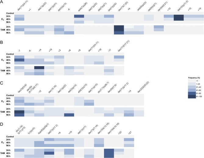
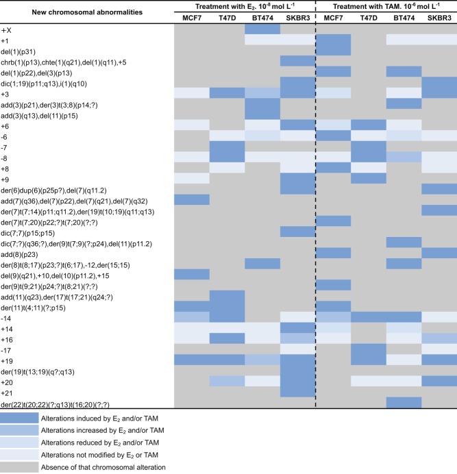
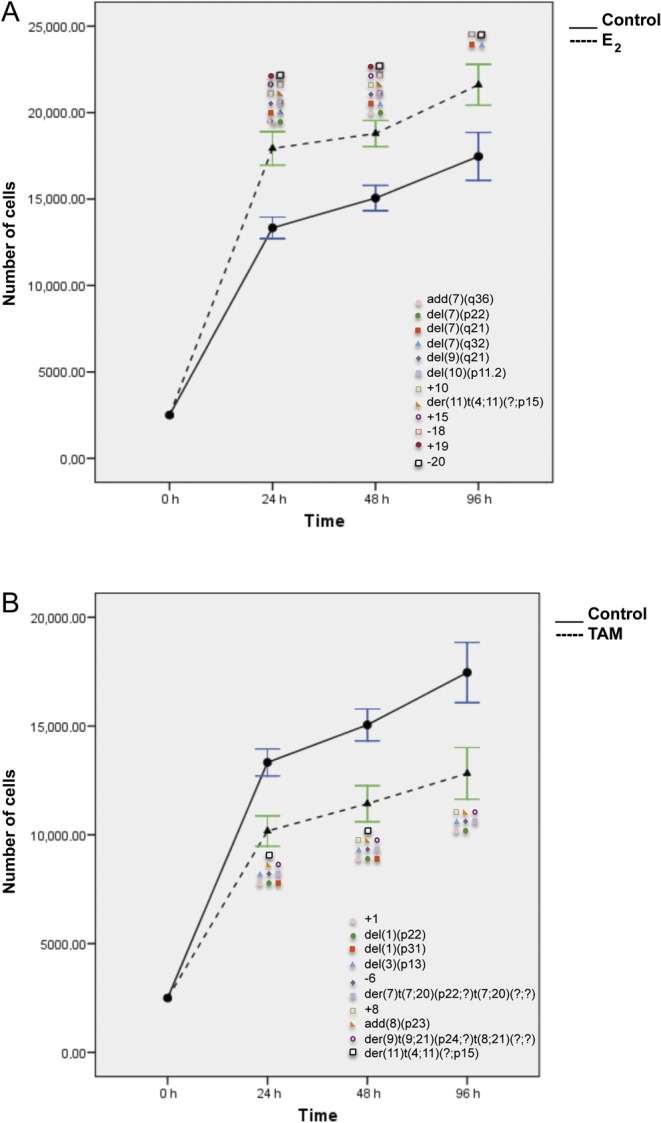
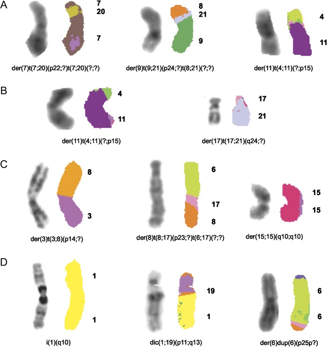
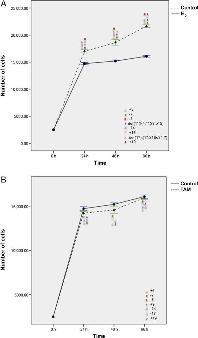
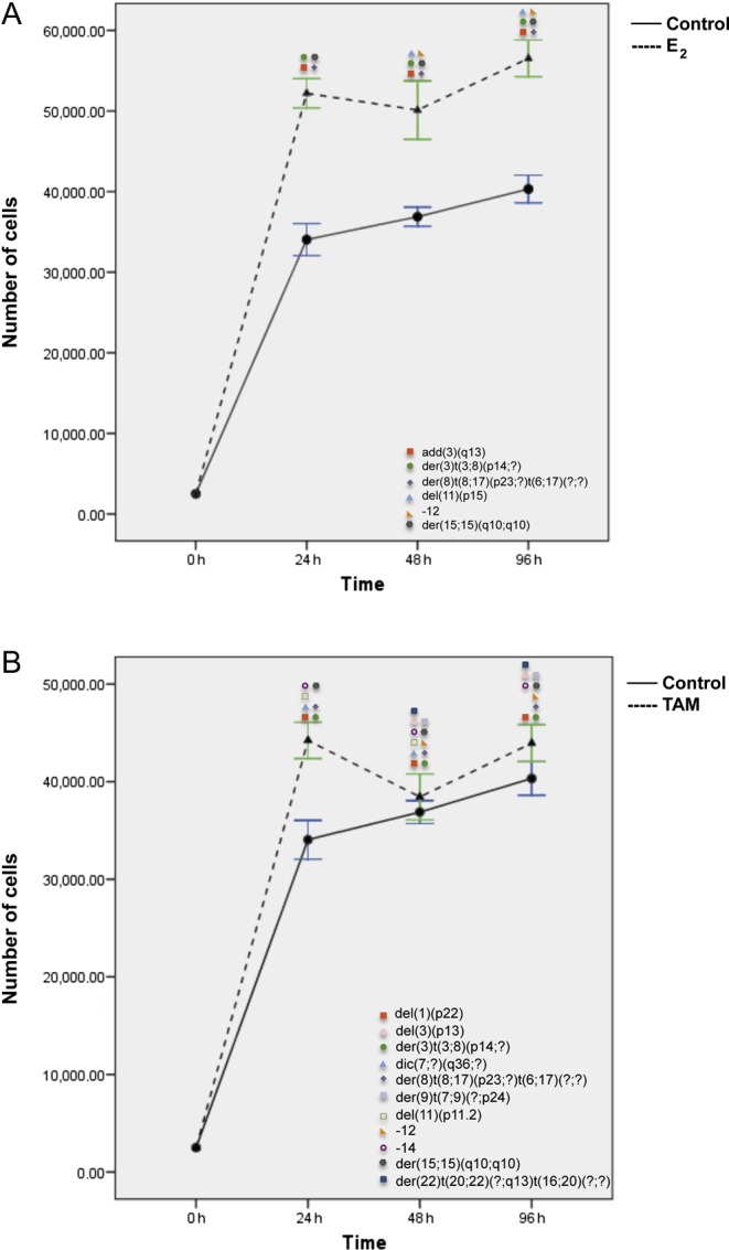
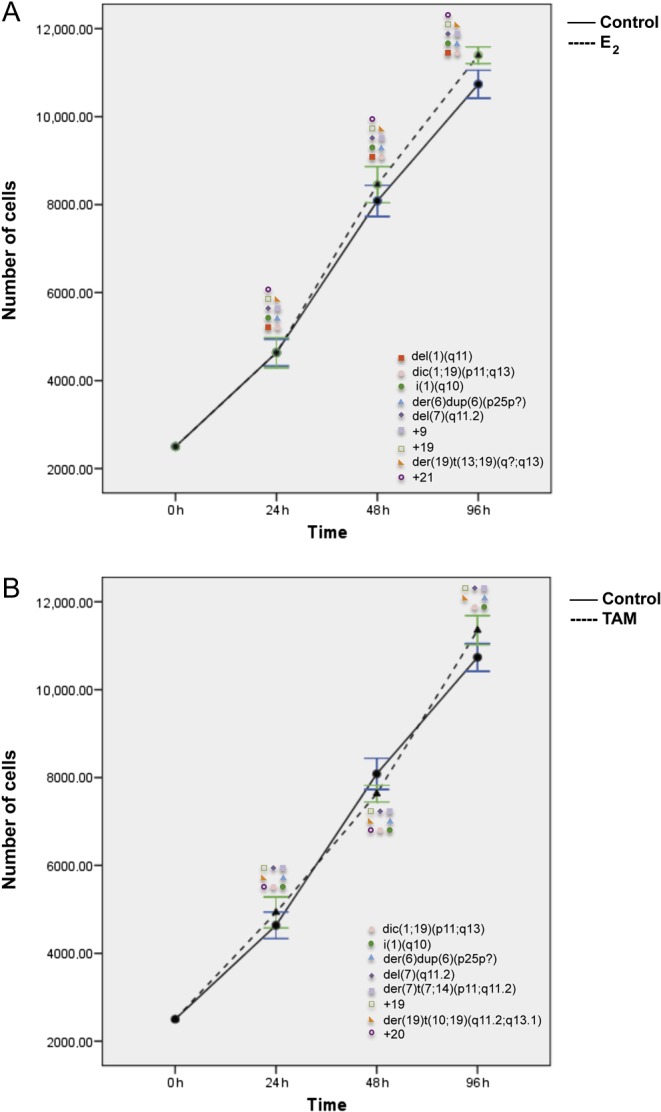
Evidence supports a role of 17&-estradiol (E2) in carcinogenesis and the large majority of breast carcinomas are dependent on estrogen. The anti-estrogen tamoxifen (TAM) is widely used for both treatment and prevention of breast cancer; however, it is also carcinogenic in human uterus and rat liver, highlighting the profound complexity of its actions. The nature of E2- or TAM-induced chromosomal damage has been explored using relatively high concentrations of these agents, and only some numerical aberrations and chromosomal breaks have been analyzed. This study aimed to determine the effects of low doses of E2 and TAM (10(&8 )mol L(&1) and 10(&6 )mol L(&1) respectively) on karyotypes of MCF7, T47D, BT474, and SKBR3 breast cancer cells by comparing the results of conventional karyotyping and multi-FISH painting with cell proliferation. Estrogen receptor (ER)-positive (+) cells showed an increase in cell proliferation after E2 treatment (MCF7, T47D, and BT474) and a decrease after TAM treatment (MCF7 and T47D), whereas in ER& cells (SKBR3), no alterations in cell proliferation were observed, except for a small increase at 96 h. Karyotypes of both ER+ and ER& breast cancer cells increased in complexity after treatments with E2 and TAM leading to specific chromosomal abnormalities, some of which were consistent throughout the treatment duration. This genotoxic effect was higher in HER2+ cells. The ER&/HER2+ SKBR3 cells were found to be sensitive to TAM, exhibiting an increase in chromosomal aberrations. These in vitro results provide insights into the potential role of low doses of E2 and TAM in inducing chromosomal rearrangements in breast cancer cells.

Keywords: breast cancer cells; chromosomal abnormalities; chromosomal instability; estradiol; tamoxifen.


Figures

Figure 1
Figure 1
Frequency of chromosomal alterations observed de novo after E2 and TAM treatments. The frequency of each chromosomal alteration is indicated along the treatments (24, 48, and 96 h) using a color code for each category. (A) MCF7 cells. (B) T47D cells. (C) BT474 cells. (D) SKBR3 cells. A full colour version of this figure is available at http://dx.doi.org/10.1530/ERC-16-0078.
Figure 2
Figure 2
Clonal chromosomal abnormalities induced by E2 and TAM in four breast cancer cell lines at each treatment time point. The presence of a given chromosomal alteration after E2 and/or TAM treatment in one or more cell lines is color coded according to the legend at the bottom. A full colour version of this figure is available at http://dx.doi.org/10.1530/ERC-16-0078.
Figure 3
Figure 3
Total number of chromosomal aberrations induced after E2 (A) and TAM (B) treatment at 24, 48, and 96 h in MCF7, T47D, BT474, and SKBR3 cell lines. Numerical chromosomal alterations: gains and losses. Structural chromosomal alterations: add, additional material of unknown origin; del, deletion; der, derivative chromosome; dic, dicentric chromosome; 
i, isochromosome. A full colour version of this figure is available at http://dx.doi.org/10.1530/ERC-16-0078.
Figure 4
Figure 4
Effects of E2 (A) and TAM (B) treatment for 24, 48, and 96 h on proliferation and corresponding chromosomal alterations in MCF7 cells. Error bars represent mean standard deviation of 24 separate experiments. Chromosomal abnormalities induced at each treatment time point are indicated. A full colour version of this figure is available at http://dx.doi.org/10.1530/ERC-16-0078.
Figure 5
Figure 5
Representative images of chromosomal abnormalities observed throughout the treatment duration with either E2 or TAM. (A) MCF7 cells, (B) T47D cells, (C) BT474 cells, and (D) SKBR3 cells. Rearranged chromosomes are visualized by G-banding technique on the left and by M-FISH on the right. The chromosomes involved in the rearrangement are numbered on the right hand side of the chromosomes. A full colour version of this figure is available at http://dx.doi.org/10.1530/ERC-16-0078.
Figure 6
Figure 6
Effects of E2 (A) and TAM (B) treatment for 24, 48, and 96 h on proliferation and corresponding chromosomal alterations in T47D cells. Error bars represent mean standard deviation of 24 separate experiments. Chromosomal abnormalities induced at each treatment time point are indicated. A full colour version of this figure is available at http://dx.doi.org/10.1530/ERC-16-0078.
Figure 7
Figure 7
Effects of E2 (A) and TAM (B) treatment for 24, 48, and 96 h on proliferation and corresponding chromosomal alterations in BT474 cells. Error bars represent mean standard deviation of 24 separate experiments. Chromosomal abnormalities induced at each treatment time point are indicated. A full colour version of this figure is available at http://dx.doi.org/10.1530/ERC-16-0078.
Figure 8
Figure 8
Effects of E2 (A) and TAM (B) treatment for 24, 48, and 96 h on proliferation and corresponding chromosomal alterations in SKBR3 cells. Error bars represent mean standard deviation of 24 separate experiments. Chromosomal abnormalities induced at each treatment time point are indicated. A full colour version of this figure is available at http://dx.doi.org/10.1530/ERC-16-0078.

References

    1. Achuthan R, Bell SM, Roberts P, Leek JP, Horgan K, Markham AF, MacLennan KA, Speirs V. 2001. Genetic events during the transformation of a tamoxifen-sensitive human breast cancer cell line into a drug-resistant clone. Cancer Genetics and Cytogenetics 130 166–172. (10.1016/S0165-4608(01)00475-7) - DOI - PubMed
    1. Ahmad ME, Shadab GG, Hoda A, Afzal M. 2000. Genotoxic effects of estradiol-17beta on human lymphocyte chromosomes. Mutation Research 466 109–115. (10.1016/S1383-5718(99)00230-2) - DOI - PubMed
    1. Al Sarraj J, Vinson C, Thiel G. 2005. Regulation of asparagine synthetase gene transcription by the basic region leucine zipper transcription factors ATF5 and CHOP. Biological Chemistry 386 873–879. (10.1515/BC.2005.102) - DOI - PubMed
    1. Azuma K, Urano T, Horie-Inoue K, Hayashi S, Sakai R, Ouchi Y, Inoue S. 2009. Association of estrogen receptor alpha and histone deacetylase 6 causes rapid deacetylation of tubulin in breast cancer cells. Cancer Research 69 2935–2940. (10.1158/0008-5472.CAN-08-3458) - DOI - PubMed
    1. Banham AH, Beasley N, Campo E, Fernandez PL, Fidler C, Gatter K, Jones M, Mason DY, Prime JE, Trougouboff P, et al. 2001. The FOXP1 winged helix transcription factor is a novel candidate tumor suppressor gene on chromosome 3p. Cancer Research 61 8820–8829. - PubMed

Publication types