Randomized Controlled Trial
. 2016 Jun 30:8:25.
doi: 10.1186/s13195-016-0194-x.

The effect of APOE genotype on the delivery of DHA to cerebrospinal fluid in Alzheimer's disease

Affiliations
Randomized Controlled Trial

The effect of APOE genotype on the delivery of DHA to cerebrospinal fluid in Alzheimer's disease

Hussein N Yassine et al. Alzheimers Res Ther. .

Abstract

Background: Apolipoprotein E (APOE) ɛ4 and low cerebrospinal fluid (CSF) amyloid-β42 (Aβ42) levels are predictors for developing Alzheimer's disease (AD). The results of several studies indicate an interaction between docosahexaenoic acid (DHA) consumption and cognitive outcomes by APOE genotype. Our objective in the present study was to examine whether APOE ɛ4 genotype and low CSF Aβ42 levels were associated with reduced delivery of DHA to CSF in the Alzheimer's Disease Cooperative Study-sponsored DHA clinical trial.

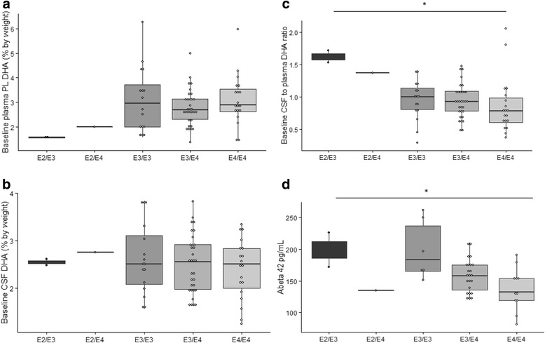
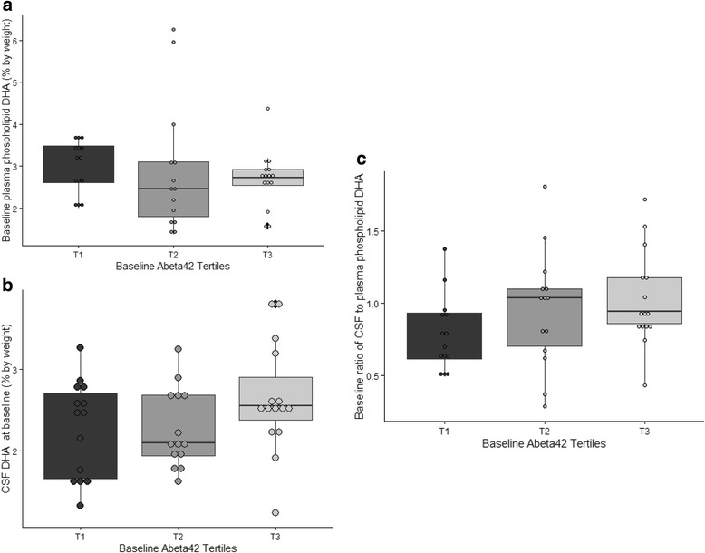
Methods: Phospholipid DHA was assayed in the plasma of 384 participants and CSF of 70 participants at baseline. Forty-four of the 70 participants completed the 18-month follow-up visit after allocation to placebo (n = 15) or DHA (n = 29). Plasma and CSF DHA levels, CSF Aβ42, Tau, and phosphorylated Tau were measured at baseline and after the 18-month intervention. Participants were divided into tertiles based on baseline Aβ42 CSF levels. To assess DHA delivery across the blood-brain barrier, the ratio of CSF to plasma DHA levels was calculated.

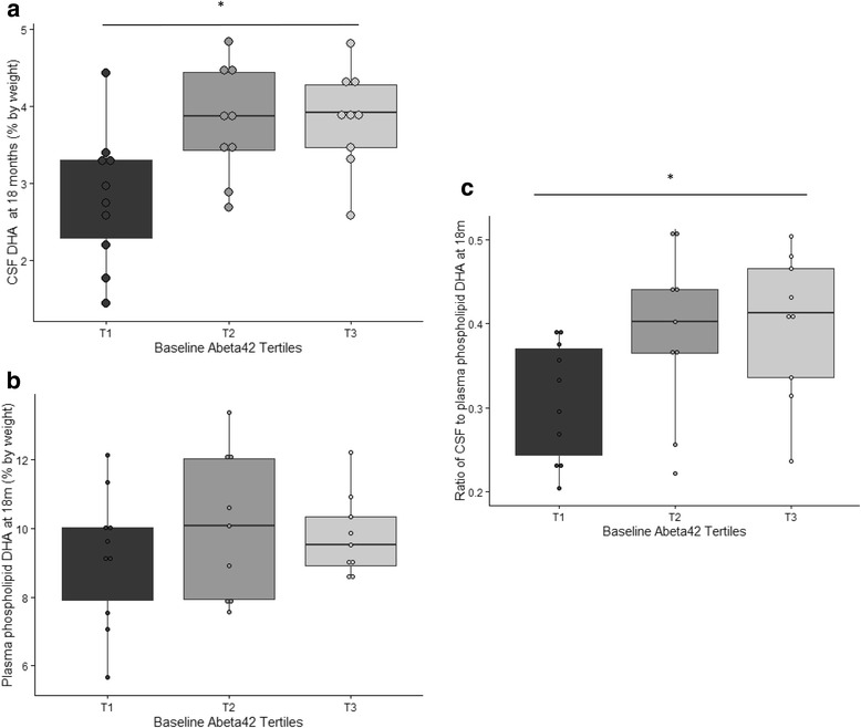
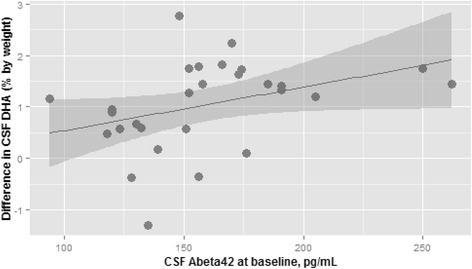
Results: At baseline, there were no significant differences between CSF or plasma phospholipid DHA levels by CSF Aβ42 tertiles or ɛ4 status. After 18 months of DHA supplementation, participants at the lowest Aβ42 tertile had significantly lower CSF DHA levels (p = 0.01) and lower CSF-to-plasma DHA ratios (p = 0.05) compared to the other tertiles. Baseline CSF Aβ42 levels were significantly lower in ɛ4 carriers than in ɛ4 noncarriers (p = 0.01). Participants carrying the ɛ4 allele (n = 25) demonstrated a less pronounced increase in CSF DHA level compared with noncarriers (n = 4), with a possible interaction effect between treatment and APOE genotype (p = 0.07).

Conclusions: APOE ɛ4 allele and lower CSF Aβ42 levels were associated with less transport of DHA to CSF. Brain amyloid pathology may limit the delivery of DHA to the brain in AD.

Trial registration: Clinicaltrials.gov identifier: NCT00440050 . Registered on 22 Feb 2007.

Keywords: APOE; Alzheimer’s disease; Amyloid; Cerebrospinal fluid.


Figures

Fig. 1
Fig. 1
Baseline CSF DHA levels by APOE status. The distributions of baseline plasma phospholipid and CSF DHA, as well as the ratio of CSF to plasma DHA, are demonstrated by APOE genotype (n = 70). There were no significant differences in (a) plasma phospholipid or (b) CSF DHA levels by APOE genotype. c The ratio of CSF to plasma DHA by APOE genotype at baseline significantly differed between the APOE genotype groups (p = 0.03 for group comparison). d CSF Aβ42 levels were significantly lower in ɛ4 carriers at baseline (p = 0.01 for group comparison). DHA levels are reported as a percentage by weight. The groups were compared using linear regression with DHA level or CSF-to-plasma DHA ratio as the dependent variable and groups as the covariate.*p < 0.05 for group comparison. Aβ42 amyloid-β42, APOE apolipoprotein E, CSF cerebrospinal fluid, DHA docosahexaenoic acid, PL Phospholipids
Fig. 2
Fig. 2
Baseline plasma phospholipid and CSF DHA levels by CSF Aβ42 tertiles. Baseline phospholipid and CSF DHA measurements were compared in three groups of participants based on CSF Aβ42 tertiles at baseline (n = 44, T1 = Aβ42 levels <147 pg/ml, T2 = Aβ42 levels between 147 and 174 pg/ml, and T3 = Aβ42 levels >174 pg/ml). The ratio of CSF to plasma DHA was calculated as an index of DHA transport across the blood-brain barrier (a and b). There was no significant difference in plasma phospholipid of CSF DHA by Aβ42 tertiles at baseline. c A trend was observed for lower baseline CSF to plasma DHA (p = 0.15 for three-way group comparison, p = 0.19 for difference between T1 and T2, and p = 0.06 for difference between T1 and T3) in the lowest tertile of Aβ42. Aβ42 amyloid-β42, CSF cerebrospinal fluid, DHA docosahexaenoic acid
Fig. 3
Fig. 3
Plasma phospholipid and CSF DHA at 18 months. The distribution of phospholipid and CSF DHA measurements at 18 months by baseline CSF Aβ42 tertiles in participants allocated to DHA (n = 29) is illustrated. a There was no significant difference in plasma phospholipid DHA by Aβ42 tertiles after 18 months of DHA. b and c After 18 months of DHA supplementation, there was a significant decrease in CSF DHA levels in participants in the the first Aβ42 tertile (p = 0.01 for three-way group comparison, p = 0.01 for difference between T1 and T2, and p = 0.007 for difference between T1 and T3) and a significantly lower increase in the CSF-to-plasma DHA ratio (p = 0.05 for three-way group comparison, p = 0.05 for difference between T1 and T2, and p = 0.03 for difference between T1 and T3). The groups were compared using a linear regression model. *p < 0.05 for group comparison. Aβ42 amyloid-β42, CSF cerebrospinal fluid, DHA docosahexaenoic acid
Fig. 4
Fig. 4
Association of CSF Aβ42 with the change in CSF DHA. Baseline CSF Aβ42 was significantly associated with the change in CSF DHA after supplementation (r = 0.37, p = 0.05). The change in DHA was calculated as the difference between 18-month CSF DHA levels and the levels at baseline. The correlation was obtained using Spearman’s correlation test. Aβ42 amyloid-β42, CSF cerebrospinal fluid, DHA docosahexaenoic acid
Fig. 5
Fig. 5
Change in CSF DHA by APOE status and treatment arm. The effect of DHA treatment vs. placebo on CSF DHA levels by APOE genotype is illustrated. The increases in DHA levels in the CSF were less pronounced in carriers of the ɛ4 allele. All ɛ4 noncarriers had increased CSF DHA levels after allocation to DHA treatment. In contrast, 6 of the 25 ɛ4 carriers did not increase DHA levels after DHA supplementation. There was a suggestion for an interaction effect between APOE genotype and treatment arm on CSF DHA levels (p = 0.07). The data were modeled using multivariate linear regression with the change in CSF DHA as the dependent variable and APOE and treatment arm as the independent variables. Aβ42 amyloid-β42, APOE apolipoprotein E, CSF cerebrospinal fluid, DHA docosahexaenoic acid

References

    1. Lambert JC, et al. Meta-analysis of 74,046 individuals identifies 11 new susceptibility loci for Alzheimer’s disease. Nat Genet. 2013;45(12):1452–8. doi: 10.1038/ng.2802. - DOI - PMC - PubMed
    1. Corder EH, et al. Gene dose of apolipoprotein E type 4 allele and the risk of Alzheimer’s disease in late onset families. Science. 1993;261(5123):921–3. doi: 10.1126/science.8346443. - DOI - PubMed
    1. Genin E, et al. APOE and Alzheimer disease: a major gene with semi-dominant inheritance. Mol Psychiatry. 2011;16(9):903–7. doi: 10.1038/mp.2011.52. - DOI - PMC - PubMed
    1. Cunnane SC, et al. Fish, docosahexaenoic acid and Alzheimer’s disease. Prog Lipid Res. 2009;48(5):239–56. doi: 10.1016/j.plipres.2009.04.001. - DOI - PubMed
    1. Cunnane SC, et al. Plasma and brain fatty acid profiles in mild cognitive impairment and Alzheimer’s disease. J Alzheimers Dis. 2012;29(3):691–7. - PMC - PubMed

Publication types

MeSH terms

Associated data

LinkOut - more resources