Case Reports
. 2016 Jun 29:2016:bcr2016215961.
doi: 10.1136/bcr-2016-215961.

Multiple brown tumours from parathyroid carcinoma

Affiliations
Case Reports

Multiple brown tumours from parathyroid carcinoma

Daryl Jade Tardo Dagang et al. BMJ Case Rep. .

Abstract

We report a case of a 29-year-old woman who suffered from severe bilateral inguinal pain and left mandibular mass. CT scan showed innumerable expansile osteolytic bone masses on the iliac wings, femur, ribs and vertebral bodies, diffuse skeletal osteopaenia, calyceal lithiasis on the right kidney and a left thyroid mass. Ionised calcium and intact parathyroid hormone (PTH) were elevated. Parathyroid sestamibi scan showed a hyperfunctioning left inferior parathyroid gland. Biopsy of the left mandibular mass was consistent with brown tumour. The patient underwent parathyroidectomy of the enlarged parathyroid gland. Final histopathology, however, revealed parathyroid carcinoma, 4.7 cm in widest dimension, with capsular and vascular space invasion. The patient underwent repeat surgery, specifically, left thyroid lobectomy, isthmectomy and central node dissection. Intact PTH decreased from 681.3 to 74 pg/mL (normal range: 10-65) 24 hours postoperatively. Follow-up at 6 months showed normal serum calcium levels, size reduction of bone lesions and improvement of quality of life.


Figures

Figure 1
Figure 1
Left mandibular mass.
Figure 2
Figure 2
Radiology findings. Plain pelvic radiograph showing ill-defined lytic lesions in bilateral iliac bones and right ischial bone (white arrows) (A). Pelvic MRI showing multiple short-TI inversion recovery hyperintense foci and focal bone expansion on iliac wings (white arrows) (B). CT scan of the pelvis, axial view, showing the lytic lesions on the femoral heads (white arrows) (C). CT scan of the abdomen, sagittal view, showing osteolytic masses on the vertebral bodies (white arrows) (D).
Figure 3
Figure 3
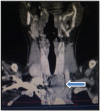
CT scan of the neck, coronal view, showing a left inferior thyroid mass (blue arrow).
Figure 4
Figure 4
Parathyroid sestamibi scan, blue arrow pointing to the hyperfunctioning gland.
Figure 5
Figure 5
H&E stain of the gingival mass showing giant cell granuloma (blue arrow) on light microscopy.
Figure 6
Figure 6
Parathyroid histology. H&E stain of parathyroid carcinoma showing capsular invasion (blue arrow) and vascular space invasion (green arrow) on light microscopy.

References

    1. Proimos E, Chimona TS, Tamiolakis D et al. . Brown tumor of the maxillary sinus in a patient with primary hyperparathyroidism: a case report. J Med Case Rep 2009;3:7495 10.4076/1752-1947-3-7495 - DOI - PMC - PubMed
    1. Belizikian JP, Silverberg SJ. Clinical Presentation of Primary Hyperparathyroidism in the United States. The Parathyroids. New York, 2001:349–60.
    1. Aoki J, Moriya K, Yamashita K et al. . Giant cell tumors of bone containing large amounts of hemosiderin: MR-pathologic correlation. J Comput Assist Tomogr 1991;15:1024–7. 10.1097/00004728-199111000-00023 - DOI - PubMed
    1. Witteveen JE, van Thiel S, Romijn JA et al. . Hungry bone syndrome: still a challenge in the post-operative management of primary hyperparathyroidism: a systematic review of the literature. Eur J Endocrinol 2013;168:R45–53. 10.1530/EJE-12-0528 - DOI - PubMed
    1. Khan A. Primary hyperparathyroidism: diagnosis and management. Endocr Pract 1997;3:22–6. 10.4158/EP.3.1.22 - DOI - PubMed

Publication types