. 2016 Aug;29(8):327-338.
doi: 10.1093/protein/gzw026. Epub 2016 Jun 29.

Probing the minimal determinants of zinc binding with computational protein design

Affiliations

Probing the minimal determinants of zinc binding with computational protein design

Sharon L Guffy et al. Protein Eng Des Sel. 2016 Aug.

Abstract

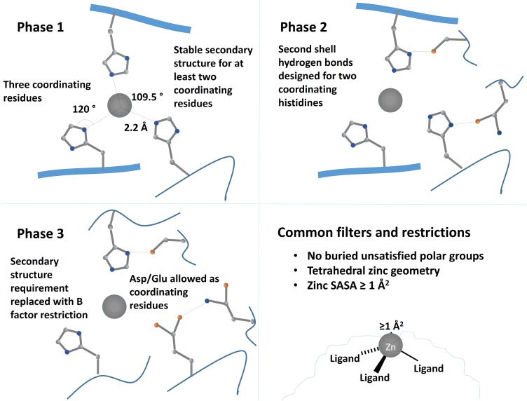
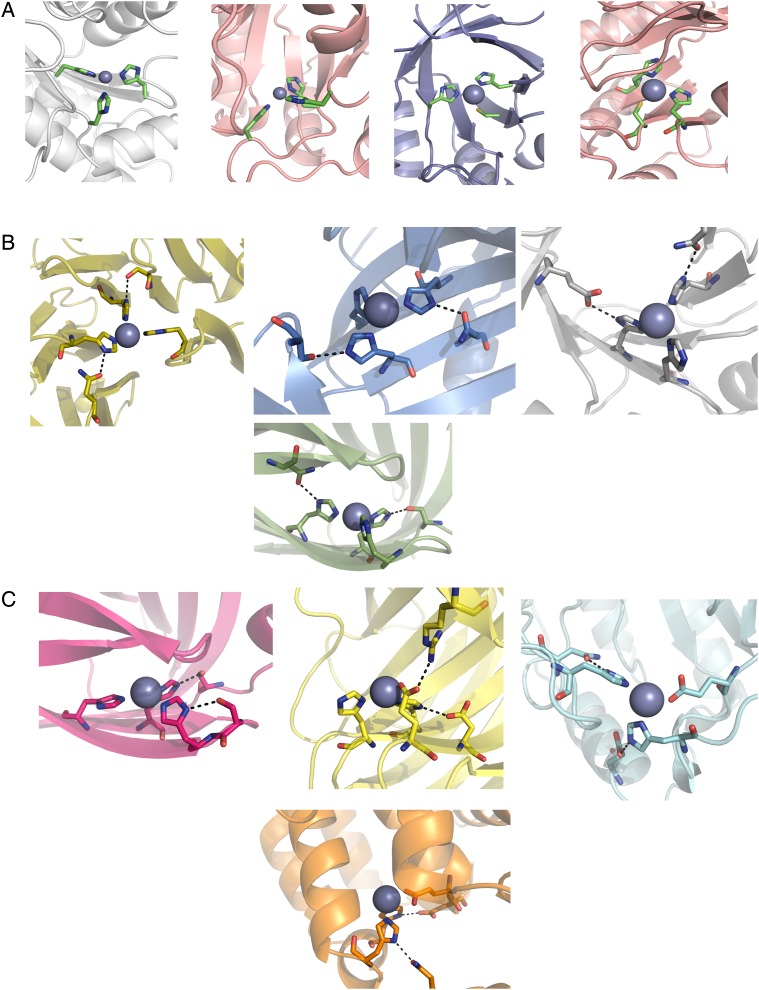
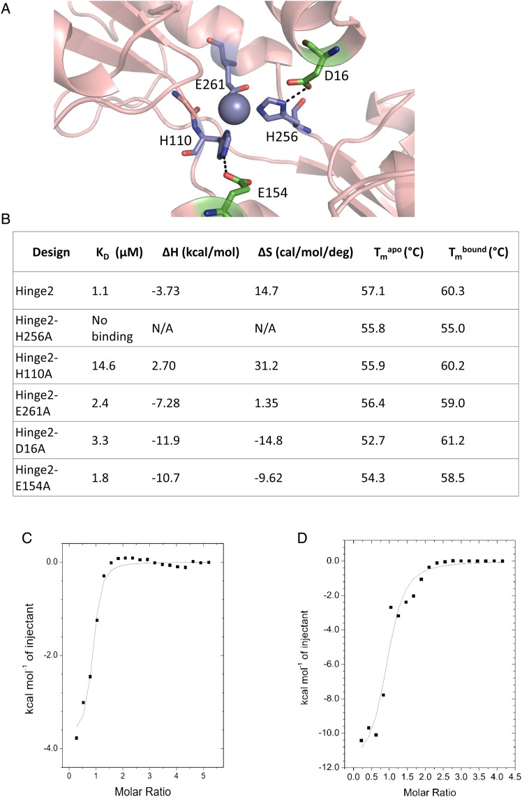
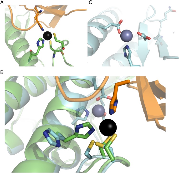
Structure-based protein design tests our understanding of the minimal determinants of protein structure and function. Previous studies have demonstrated that placing zinc binding amino acids (His, Glu, Asp or Cys) near each other in a folded protein in an arrangement predicted to be tetrahedral is often sufficient to achieve binding to zinc. However, few designs have been characterized with high-resolution structures. Here, we use X-ray crystallography, binding studies and mutation analysis to evaluate three alternative strategies for designing zinc binding sites with the molecular modeling program Rosetta. While several of the designs were observed to bind zinc, crystal structures of two designs reveal binding configurations that differ from the design model. In both cases, the modeling did not accurately capture the presence or absence of second-shell hydrogen bonds critical in determining binding-site structure. Efforts to more explicitly design second-shell hydrogen bonds were largely unsuccessful as evidenced by mutation analysis and low expression of proteins engineered with extensive primary and secondary networks. Our results suggest that improved methods for designing interaction networks will be needed for creating metal binding sites with high accuracy.

Keywords: Rosetta; metalloproteins; protein design; zinc binding.


Figures

Fig. 1
Fig. 1
Schematic of design strategies for first, second and third rounds of design. In the first set of designs (Phase 1), tetrahedral zinc binding sites were designed with three coordinating residues and one open coordination site with at least two of the ligands on stable secondary-structure elements. In Phase 2, we designed two second-shell hydrogen bonding residues in addition to three coordinating histidines. To increase the diversity of designed sites, Phase 3 allowed one of the coordinating residues to be an aspartate or glutamate and removed the secondary-structure requirement for ligand residues, instead requiring all ligand residues to have Cα B factor values of 30 or greater.
Fig. 2
Fig. 2
(A) Predicted structure of ZE2 zinc binding site. (B) Crystal structure of ZE2 zinc binding site. (C) Crystal structure of ZE2 overlaid with the design model. Although the predicted residues do form the zinc binding site, the histidines (along with the zinc ion) have shifted their positions to form stabilizing hydrogen bonds with surrounding residues. H210 (bottom center) has changed rotamers to form a hydrogen bond with the side chain of E50; H179 (left) has changed rotamers to form a hydrogen bond with N160 and is coordinating the zinc ion with the epsilon nitrogen instead of the delta nitrogen; and the loop containing H183 (top center, far right) has moved over 9 Å to accommodate the new position of the binding site. (D) ZE2 is well folded in the absence of zinc. (E) ZE2 shows increased thermal stability in the presence of saturating zinc, which increases the thermal transition temperature from 90.2 to 92.4°C. (F) ZE2 binds zinc in the designed site with 1.4 µM affinity as measured by ITC. A second nonspecific binding event (KD = 92 µM) is also observed by ITC, but only the first site appears in the crystal structure.
Fig. 3
Fig. 3
Sample models of designed zinc binding sites showing coordinating residues and hydrogen bonding residues. (A) Phase 1 sites include three histidines or cysteines with no designed second-shell residues. (B) Phase 2 sites add two second-shell hydrogen bonding residues. (C) Phase 3 sites introduce liganding Asp/Glu and relaxed secondary-structure requirements to increase site variability.
Fig. 4
Fig. 4
(A) Design model for Hinge2. The predicted binding site is composed of two histidines and one aspartate. Stabilizing hydrogen bonds were designed for two of the residues. (B) Comparison of zinc binding measurements for Hinge2 and point mutants of its three zinc-coordinating residues (H256A, H110A, E261A) and two hydrogen bond partners for those residues (D16A, E154A). All mutants show decreased affinity for zinc. The H256A mutation abolishes zinc binding. N/A, not applicable. (C and D) Sample ITC data for Hinge2 (C) and its D16A mutant (D).
Fig. 5
Fig. 5
Crystal structure of Spelter (Der et al., 2013) compared with design model. (A) The modeled zinc binding site includes two cysteines and a histidine from Spelter in addition to a residue from ubiquitin. (B) Overlay of design model and crystal structure. The designed zinc binding site (foreground) does not form. (C) An additional unmodeled zinc binding site was formed due to a mutation (V122E) made to increase stability during the design process.

Similar articles

Cited by

References

    1. Andreini C., Bertini I. (2012) J. Inorg. Biochem., 111, 150–156. - PubMed
    1. Andreini C., Bertini I., Cavallaro G., Holliday G.L., Thornton J.M. (2008) J. Biol. Inorg. Chem., 13, 1205–1218. - PubMed
    1. Auld D.S. (2001) Biometals, 14, 271–313. - PubMed
    1. Azia A., Levy R., Unger R., Edelman M., Sobolev V. (2015) Proteins Struct. Funct. Bioinforma., 83, 931–939. - PubMed
    1. Browner M.F., Hackos D., Fletterick R. (1994) Nat. Struct. Mol. Biol., 1, 327–333. - PubMed

Publication types

LinkOut - more resources