. 2016 Sep 1;116(3):1161-74.
doi: 10.1152/jn.00305.2016. Epub 2016 Jun 29.

Functional evidence for a direct excitatory projection from the lateral habenula to the ventral tegmental area in the rat

Affiliations

Functional evidence for a direct excitatory projection from the lateral habenula to the ventral tegmental area in the rat

P Leon Brown et al. J Neurophysiol. .

Abstract

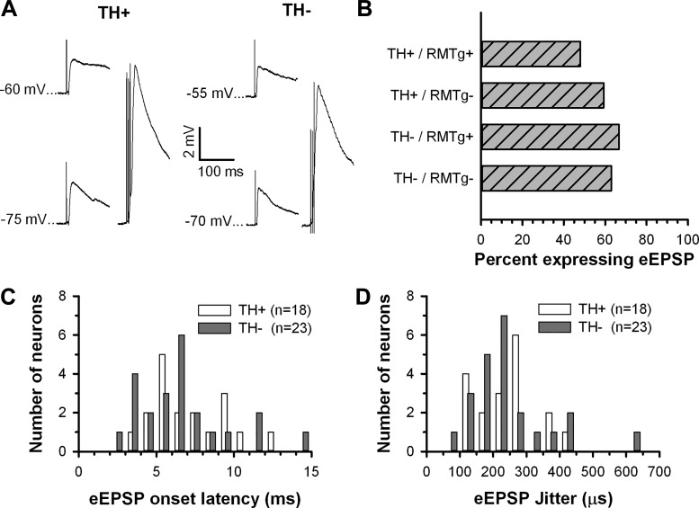
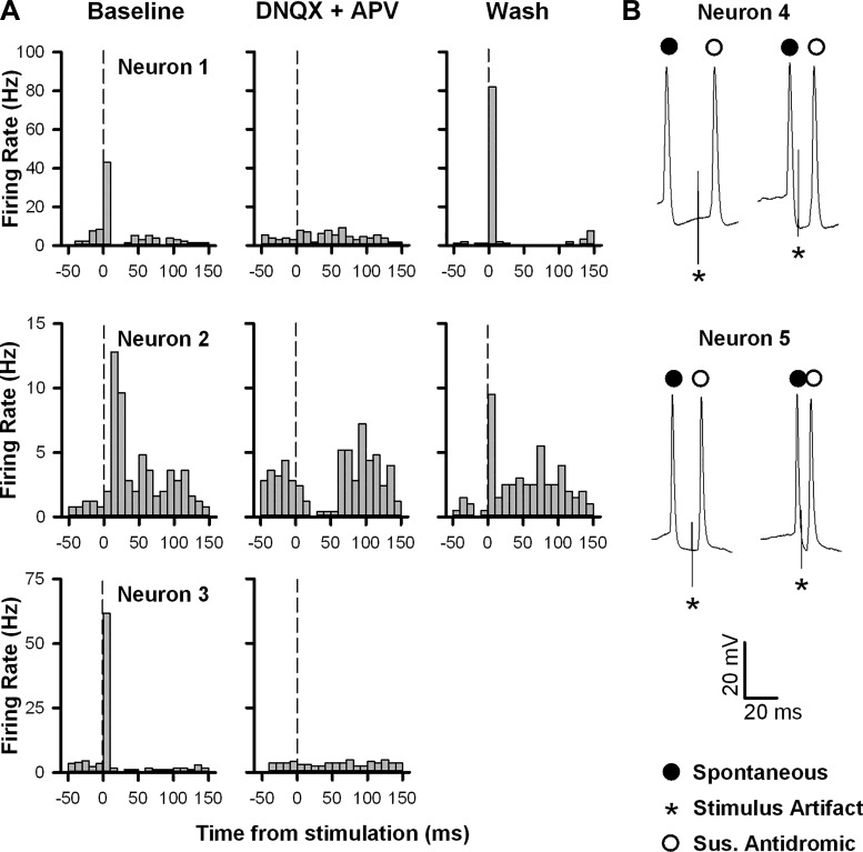
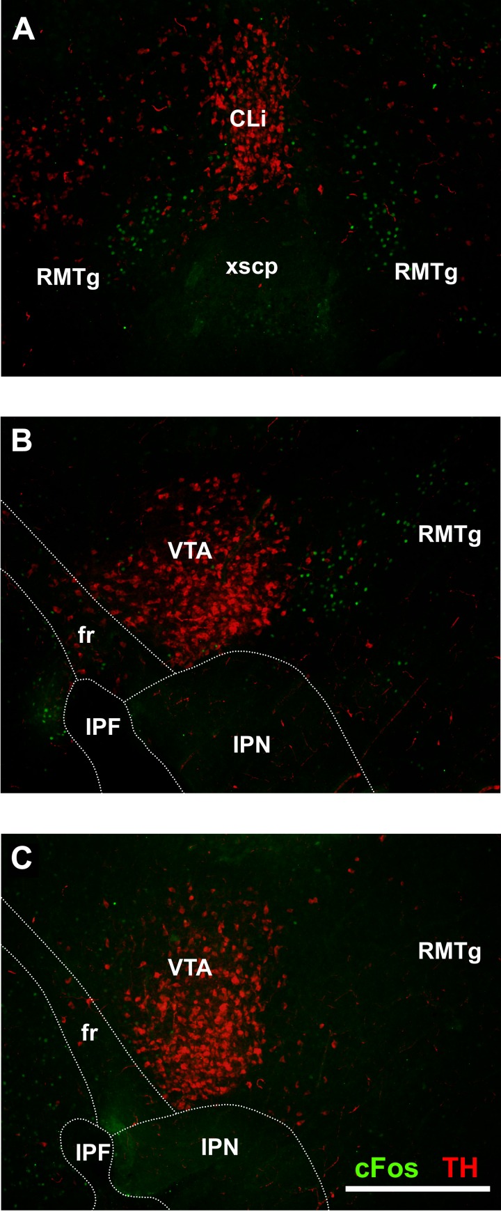
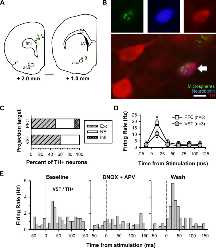
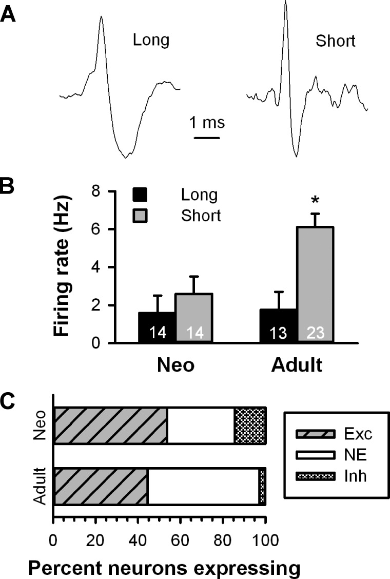
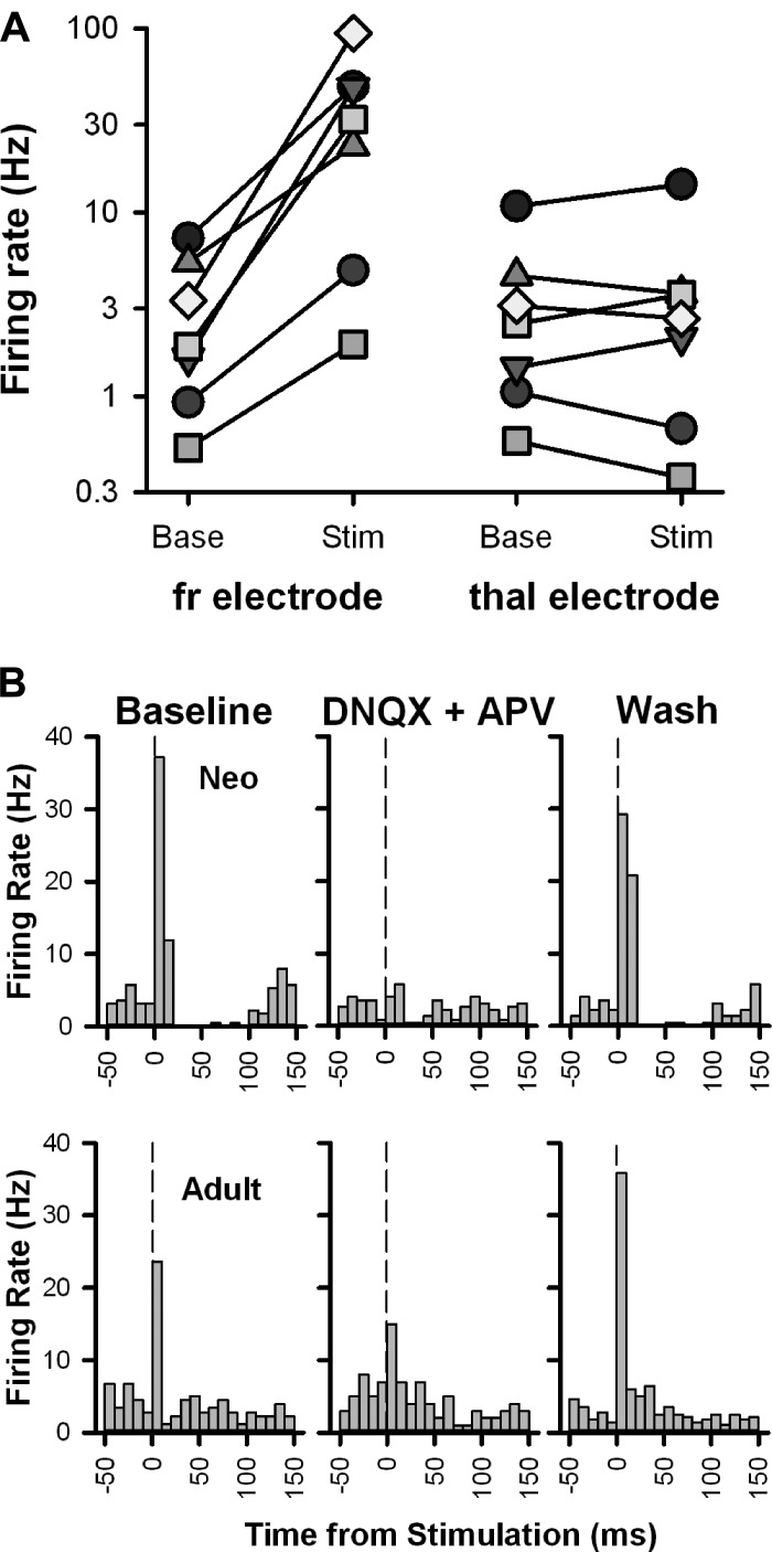
The lateral habenula, a phylogenetically conserved epithalamic structure, is activated by aversive stimuli and reward omission. Excitatory efferents from the lateral habenula predominately inhibit midbrain dopamine neuronal firing through a disynaptic, feedforward inhibitory mechanism involving the rostromedial tegmental nucleus. However, the lateral habenula also directly targets dopamine neurons within the ventral tegmental area, suggesting that opposing actions may result from increased lateral habenula activity. In the present study, we tested the effect of habenular efferent stimulation on dopamine and nondopamine neurons in the ventral tegmental area of Sprague-Dawley rats using a parasagittal brain slice preparation. Single pulse stimulation of the fasciculus retroflexus excited 48% of dopamine neurons and 51% of nondopamine neurons in the ventral tegmental area of rat pups. These proportions were not altered by excision of the rostromedial tegmental nucleus and were evident in both cortical- and striatal-projecting dopamine neurons. Glutamate receptor antagonists blocked this excitation, and fasciculus retroflexus stimulation elicited evoked excitatory postsynaptic potentials with a nearly constant onset latency, indicative of a monosynaptic, glutamatergic connection. Comparison of responses in rat pups and young adults showed no significant difference in the proportion of neurons excited by fasciculus retroflexus stimulation. Our data indicate that the well-known, indirect inhibitory effect of lateral habenula activation on midbrain dopamine neurons is complemented by a significant, direct excitatory effect. This pathway may contribute to the role of midbrain dopamine neurons in processing aversive stimuli and salience.

Keywords: brain slice; dopamine; lateral habenula; rostromedial tegmental nucleus; tail of the ventral tegmental area; tyrosine hydroxylase.


Figures

Fig. 1.
Fig. 1.
Electrophysiological properties of immunohistochemically identified ventral tegmental area (VTA) neurons. A: photomicrographs of neurobiotin-filled cells (green) in brain slices processed for tyrosine hydroxylase (TH) immunoreactivity (ir; red). The white arrow indicates the soma of a neurobiotin-filled TH+ cell. Scale bar = 50 μm. B and C: spontaneously firing neurons. TH+ VTA neurons tended to fire more slowly and exhibit wider spikes than TH VTA neurons.
Fig. 2.
Fig. 2.
Distribution of spontaneous firing rates (A), spike durations (B), and time constant (tau; C) obtained from TH+ and TH neurons recorded in the VTA.
Fig. 3.
Fig. 3.
Representative examples of the effect of fasciculus retroflexus (fr) stimulation on the activity of TH+ VTA neurons. A and B: peristimulus time histograms (PSTHs) illustrating the response of two VTA dopamine neurons to single pulse stimulation of the fr. The dashed vertical line denotes the onset of the stimulus pulse. C and D: corresponding CUMSUM plots were compiled from a running sum of the bin totals comprising the PSTHs. The dashed lines illustrate the slope of a least-squares regression line fit to each segment of the CUMSUM plot to determine the duration and magnitude of firing rate changes in response to fr stimulation (see materials and methods for details).
Fig. 4.
Fig. 4.
Summary of the effects of fr stimulation on the activity of VTA neurons. A: bar histogram illustrating the distribution of responses to fr stimulation among TH+ and TH neurons in slices with and without the rostromedial tegmental nucleus (RMTg+ and RMTg, respectively). B: the latency to onset (left axis) and duration of fr-induced excitation (right axis) were longer in TH+ neurons than in TH neurons. C: plot of the time course of spontaneous activity showing the rapid excitation and return to baseline after fr stimulation. *Group difference (P < 0.05) in B and C.
Fig. 5.
Fig. 5.
Summary of the effects of fr stimulation on synaptic potentials recorded in VTA neurons. A: fr stimulation elicited evoked excitatory postsynaptic potentials (eEPSPs) in both TH+ (left) and TH (right) VTA neurons that increased in amplitude during membrane hyperpolarization (bottom left in each example) and showed evidence of temporal summation (bottom right in each example, 3 pulses at 50 Hz). B: bar histogram illustrating the frequency of occurrence of eEPSPs in TH+ and TH neurons in slices with and without the RMTg. C and D: neither latency to EPSP onset nor SD of onset latency (jitter) differed between TH+ and TH VTA neurons.
Fig. 6.
Fig. 6.
fr stimulation induced excitation of VTA neurons is glutamate receptor dependent. A: PSTHs illustrating the changes in firing rate elicited by fr stimulation in three separate neurons before (left), during (middle), and after (right) bath application of 6,7-dinitroquinoxaline-2,3-dione (DNQX; 10 μM) and (2R)-amino-5-phosphonovaleric acid (APV; 50 μM). Vertical lines denote the stimulus onset. B: naturally occurring collision tests were evaluated in two neurons with a rapid, high-fidelity response to fr stimulation. Shown for each neuron are spontaneous spikes (closed circles) with a delay to stimulation (*) that was either longer (left trace) or shorter (right trace) than the onset latency to the suspected antidromic spike (open circle). In both neurons, short delays to stimulation failed to produce collisions, demonstrating that the activation was not antidromic. All five neurons in this figure were subsequently shown to be TH.
Fig. 7.
Fig. 7.
Photomicrographs of neonatal rat brain sections immunostained for TH (red) and c-Fos (green) after administration of (+)-methamphetamine hydrochloride. A: coronal section (150 μm) showing c-Fos+ cells ventral and lateral to the TH+ caudal linear nucleus. This c-Fos+ area corresponds to the RMTg in adult rats. B: parasagittal section (100 μm, but otherwise identical to that used in the recording experiments) showing c-Fos+ cells posterior to the TH+ VTA. C: comparable section from a vehicle-treated rat showing TH+ neurons in the VTA but no evidence of c-Fos+ neurons in the RMTg. CLi, caudal linear nucleus; IPF, interpeduncular fossa; IPN, interpeduncular nucleus; xscP, decussation of the superior cerebellar peduncle. Scale bar = 500 μm.
Fig. 8.
Fig. 8.
Use of retrograde-traveling fluorescent microspheres to label and record from projection-specific neurons. A: location of microsphere injection sites in the prefrontal cortex (PFC; left) and ventral striatum (VST; right) with approximate distance from the bregma for each. The black circle indicates an injection site ∼0.5 mm posterior to the coronal section shown. Scale bar = 1 mm. aca, Anterior commissure, anterior; cc, corpus collosum; fmi, forceps minor corpus collosum; LV, lateral ventricle; rf, rhinal fissure. B: photomicrographs of a microsphere-filled cell (top left, green) labeled with neurobiotin (top middle, blue) and stained for TH (top right, red). Merge of photomicrographs shows the recorded neuron (bottom, white arrow). Scale bar = 10 μm. C: bar histogram illustrating the distribution of responses to fr stimulation between PFC- and VST-projecting TH+ neurons. D: plot of the time course of spontaneous activity showing the rapid excitation that was stronger in PFC- than VST-projecting neurons. *Group difference (P < 0.05). E: PSTHs illustrating the changes in firing rate elicited by fr stimulation in a VST-projecting TH+ neuron before (left), during (middle), and after (right) bath application of DNQX (10 μM) and APV (50 μM). The vertical lines denote the stimulus onset.
Fig. 9.
Fig. 9.
Comparison of extracellular single unit recordings of VTA neurons from neonatal and adult parasagittal slices. A: most spontaneously firing VTA neurons from both neonatal and adult slices presented either a long-duration (left) or short-duration waveform (right). B: bar histogram illustrating the firing rate of long- and short-duration waveform neurons in neonatal and adult VTA neurons. *Group difference within short waveform neurons (P < 0.05). C: bar histogram illustrating the distribution of responses to fr stimulation between neonatal and adult VTA neurons.
Fig. 10.
Fig. 10.
Stimulation-induced excitation of VTA neurons is fr specific and glutamate receptor dependent. A: seven neurons (each represented by a different symbol) that responded with excitation to fr stimulation (left) were also tested for their response to thalamic stimulation (right). While there was a strong increase in firing rate from baseline after fr stimulation in all cells, there was no consistent change in firing rate after thalamic stimulation. Note that the ordinate is a log scale to make changes in neurons with slow firing rates more visible. B: PSTHs illustrating the changes in firing rate elicited by fr stimulation in neurons from a neonate and adult rat before (left), during (middle), and after (right) bath application of DNQX (10 μM) and APV (50 μM). The vertical lines denote the stimulus onset.

Similar articles

Cited by

References

    1. Aghajanian GK, Rasmussen K. Intracellular studies in the facial nucleus illustrating a simple new methd for obtaining viable motoneurons in adult rat brain slices. Synapse 3: 331–338, 1989. - PubMed
    1. Balcita-Pedicino JJ, Omelchenko N, Bell R, Sesack SR. The inhibitory influence of the lateral habenula on midbrain dopamine cells: Ultrastructural evidence for indirect mediation via the rostromedial mesopontine tegmental nucleus. J Comp Neurol 519: 1143–1164, 2011. - PMC - PubMed
    1. Benabid AL, Jeaugey L. Cells of the rat lateral habenula respond to high-threshold somatosensory inputs. Neurosci Lett 96: 289–294, 1989. - PubMed
    1. Blanchard V, Raisman-Vozari R, Vyas S, Michel PP, Javoy-Agid F, Uhl G, Agid Y. Differential expression of tyrosine hydroxylase and membrane dopamine transporter genes in subpopulations of dopaminergic neurons of the rat mesencephalon. Brain Res Mol Brain Res 22: 29–38, 1994. - PubMed
    1. Brinschwitz K, Dittgen A, Madai VI, Lommel R, Geisler S, Veh RW. Glutamatergic axons from the lateral habenula mainly terminate on GABAergic neurons of the ventral midbrain. Neuroscience 168: 463–476, 2010. - PubMed

Publication types

MeSH terms

LinkOut - more resources