. 2016 Aug 19;291(34):17496-17509.
doi: 10.1074/jbc.M116.740308. Epub 2016 Jun 29.

Gadd45a Protein Promotes Skeletal Muscle Atrophy by Forming a Complex with the Protein Kinase MEKK4

Affiliations

Gadd45a Protein Promotes Skeletal Muscle Atrophy by Forming a Complex with the Protein Kinase MEKK4

Steven A Bullard et al. J Biol Chem. .

Abstract

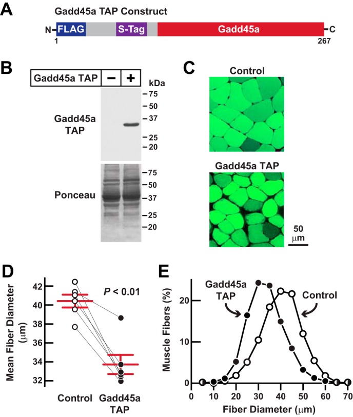
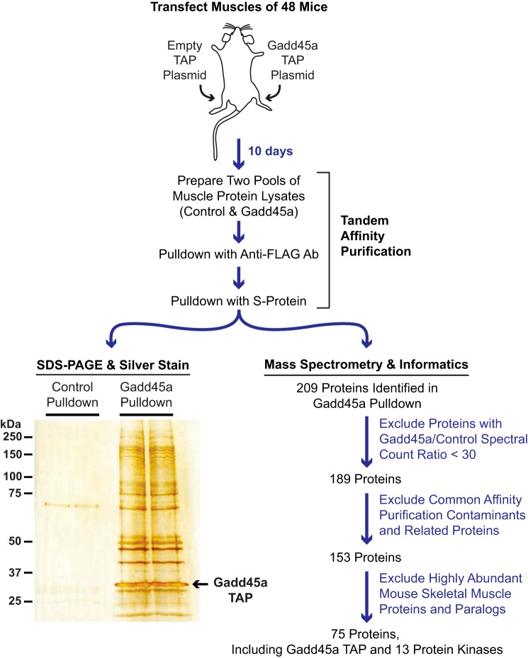
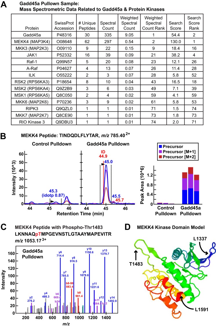
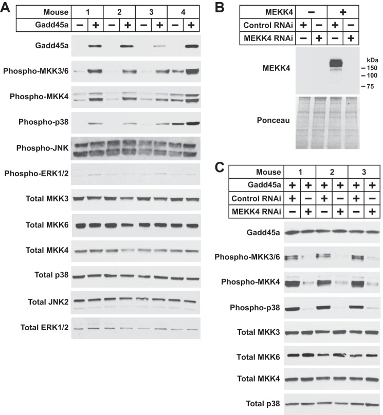
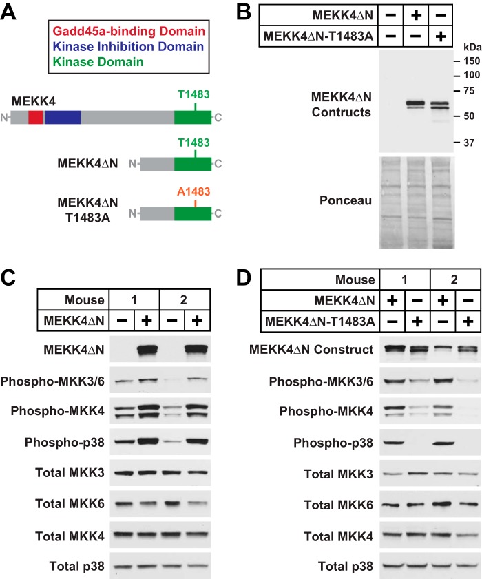
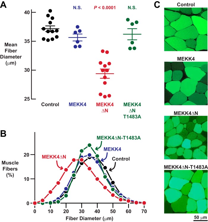
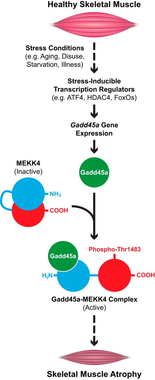
Skeletal muscle atrophy is a serious and highly prevalent condition that remains poorly understood at the molecular level. Previous work found that skeletal muscle atrophy involves an increase in skeletal muscle Gadd45a expression, which is necessary and sufficient for skeletal muscle fiber atrophy. However, the direct mechanism by which Gadd45a promotes skeletal muscle atrophy was unknown. To address this question, we biochemically isolated skeletal muscle proteins that associate with Gadd45a as it induces atrophy in mouse skeletal muscle fibers in vivo We found that Gadd45a interacts with multiple proteins in skeletal muscle fibers, including, most prominently, MEKK4, a mitogen-activated protein kinase kinase kinase that was not previously known to play a role in skeletal muscle atrophy. Furthermore, we found that, by forming a complex with MEKK4 in skeletal muscle fibers, Gadd45a increases MEKK4 protein kinase activity, which is both sufficient to induce skeletal muscle fiber atrophy and required for Gadd45a-mediated skeletal muscle fiber atrophy. Together, these results identify a direct biochemical mechanism by which Gadd45a induces skeletal muscle atrophy and provide new insight into the way that skeletal muscle atrophy occurs at the molecular level.

Keywords: Gadd45a; MEKK4; aging; mass spectrometry (MS); muscle; muscle atrophy; skeletal muscle; skeletal muscle metabolism.


Figures

FIGURE 1.
FIGURE 1.
Gadd45a TAP, a functional Gadd45a construct designed for tandem affinity purification in mouse skeletal muscle. A, schematic of the Gadd45a TAP construct. B–E, mouse TA muscle fibers were transfected with 20 μg of Gadd45a TAP plasmid plus 2.5 μg of eGFP plasmid. In each mouse, the contralateral TA muscle fibers (Control) were transfected with 20 μg of empty TAP plasmid plus 2.5 μg of eGFP plasmid. Bilateral TA muscles were harvested for analysis 7 days post-transfection. B, skeletal muscle protein extracts were subjected to immunoblot analysis using monoclonal anti-FLAG IgG. C, representative fluorescence microscopy images of muscle cross-sections. D, average diameters of skeletal muscle fibers. Each data point represents the mean of >450 muscle fibers from one muscle, and horizontal bars denote average of the means ± S.E. p value was determined with a paired t test. E, size distribution of all muscle fibers from D.
FIGURE 2.
FIGURE 2.
Isolation and analysis of proteins that interact with Gadd45a TAP in mouse skeletal muscle fibers. TA skeletal muscle fibers of 48 mice were transfected with 20 μg of empty TAP plasmid (one TA per mouse) or 20 μg of Gadd45a TAP plasmid (the contralateral TA in each mouse). Ten days post-transfection, bilateral TA muscles were harvested and used to prepare pooled protein extracts from each of the two groups of skeletal muscles (control and Gadd45a). The pooled protein extracts were then subjected to sequential purification steps with anti-FLAG magnetic beads and S-protein affinity gel. An aliquot of each final pulldown sample was visualized by SDS-PAGE and silver staining, as shown. The remaining portions of control and Gadd45a pulldown samples were subjected to mass spectrometry and a data analysis workflow that is summarized here and fully detailed under “Experimental Procedures” and the supplemental Tables.
FIGURE 3.
FIGURE 3.
Gadd45a TAP interacts with multiple protein kinases in mouse skeletal muscle, including MEKK4. A, summary of mass spectroscopic data for Gadd45a and associated protein kinases in the Gadd45a pulldown sample. B, data from a representative MEKK4 peptide (TINDQDLFLYTAR with the precursor ion at m/z 785.402+) identified in the Gadd45a pulldown sample. Left and middle panels, quantification using MS1 filtering; the panels show extracted ion chromatograms for the MS1 precursor ions in the control and Gadd45a pulldown samples. Right panel, calculated peak areas under the curve for three technical replicates of the control and Gadd45a pulldown samples. C, MS/MS of MEKK4 phosphopeptide in the Gadd45a pulldown sample. The b8 ion (m/z 981.4) and the corresponding b8-98 ion (m/z 883.4), resulting from a neutral loss of H3PO4 (−98), clearly indicates that phosphorylation in this peptide is located at threonine 1483. D, three-dimensional model of the mouse MEKK4 kinase domain (Sprot number O08648, residues 1337–1591), indicating the position of threonine 1483 within the activation loop. The model, generated by The Protein Model Portal, is based on the crystal structure of the Mst1 kinase (template 3comA, sequence identity 33%).
FIGURE 4.
FIGURE 4.
Gadd45a activates MEKK4 in mouse skeletal muscle. A, one TA per mouse was transfected with 10 μg of empty pcDNA plasmid, and the contralateral TA in each mouse was transfected with 10 μg of Gadd45a-FLAG plasmid, as indicated. Seven days post-transfection, bilateral TA muscles were harvested for immunoblot analysis using the indicated antibodies. B, mouse TA muscles were transfected with 5 μg of empty pcDNA plasmid, 5 μg of MEKK4-FLAG plasmid, 20 μg of control RNAi plasmid, and/or 20 μg of MEKK4 RNAi plasmid, as indicated. Seven days post-transfection, TA muscles were harvested for immunoblot analysis using an anti-FLAG antibody. C, one TA per mouse was transfected with 10 μg of Gadd45a-FLAG plasmid plus 20 μg of control RNAi plasmid, and the contralateral TA in each mouse was transfected with 10 μg of Gadd45a-FLAG plasmid plus 20 μg of MEKK4 RNAi plasmid, as indicated. Seven days post-transfection, bilateral TA muscles were harvested for immunoblot analysis using the indicated antibodies.
FIGURE 5.
FIGURE 5.
MEKK4 is required for Gadd45a-mediated skeletal muscle fiber atrophy. A–C, in one cohort of mice, one TA per mouse was transfected with 20 μg of control RNAi plasmid, and the contralateral TA in each mouse was transfected with 20 μg of MEKK4 RNAi plasmid. In a second cohort of mice, one TA per mouse was transfected with 10 μg of Gadd45a-FLAG plasmid plus 20 μg of control RNAi plasmid, and the contralateral TA in each mouse was transfected with 10 μg of Gadd45a-FLAG plasmid plus 20 μg of MEKK4 RNAi plasmid. In all mice, bilateral TA muscles were harvested for histological analysis 7 days post-transfection. A, average diameters of skeletal muscle fibers. Each data point represents the mean of >500 muscle fibers from one muscle, and horizontal bars denote average of the means ± S.E. p values were determined with paired t tests. N.S., not significant or p > 0.05. B, size distribution of all muscle fibers from A. C, representative fluorescence microscopy images of muscle cross-sections. D, in one cohort of mice, bilateral TA muscles were transfected with 20 μg of control RNAi plasmid. In a second cohort of mice, bilateral TA muscles were transfected with 20 μg of MEKK4 RNAi plasmid. Three days after transfection, one hindlimb in each mouse was immobilized, and then 7 days later (10 days post-transfection), bilateral TA muscles were harvested for histological analysis. In each mouse, the average muscle fiber diameter in the immobilized TA was normalized to the average muscle fiber diameter in the contralateral control (mobile) TA. Each data point represents the value from one mouse, and horizontal bars denote averages ± S.E.
FIGURE 6.
FIGURE 6.
MEKK4ΔN, a constitutively active Gadd45a-independent MEKK4 construct. A, schematic of the MEKK4ΔN and MEKK4ΔN-T1483A constructs. B, TA muscles were transfected with 5 μg of empty pcDNA plasmid, 5 μg of MEKK4ΔN-FLAG plasmid, or 5 μg of MEKK4ΔN-T1483A-FLAG plasmid, as indicated. Three days post-transfection, TA muscles were harvested for immunoblot analysis using monoclonal anti-FLAG IgG. C, one TA per mouse was transfected with 5 μg of empty pcDNA plasmid, and the contralateral TA in each mouse was transfected with 5 μg of MEKK4ΔN-FLAG plasmid, as indicated. Three days post-transfection, bilateral TA muscles were harvested for immunoblot analysis using the indicated antibodies. D, one TA per mouse was transfected with 5 μg of MEKK4ΔN-FLAG plasmid, and the contralateral TA in each mouse was transfected with 5 μg of MEKK4ΔN-T1483A-FLAG plasmid, as indicated. Three days post-transfection, bilateral TA muscles were harvested for immunoblot analysis using the indicated antibodies.
FIGURE 7.
FIGURE 7.
MEKK4ΔN induces skeletal muscle fiber atrophy in a Gadd45a-independent manner. A–C, mouse TA muscles were transfected with 5 μg of empty pcDNA plasmid, 5 μg of MEKK4-FLAG plasmid, 5 μg of MEKK4ΔN-FLAG plasmid, and/or 5 μg of MEKK4ΔN-T1483A-FLAG plasmid, as indicated. All muscles were co-transfected with 2.5 μg of eGFP plasmid. TA muscles were harvested for histological analysis 7 days post-transfection. A, average diameters of skeletal muscle fibers. Each data point represents the mean of >400 muscle fibers from one muscle, and horizontal bars denote average of the means ± S.E. p values were determined with a one-way ANOVA and Dunnett's multiple comparison test. N.S., not significant or p > 0.05. B, size distribution of all muscle fibers from A. C, representative fluorescence microscopy images of muscle cross-sections.
FIGURE 8.
FIGURE 8.
Proposed model illustrating the role of MEKK4 in Gadd45a-mediated skeletal muscle atrophy.

Similar articles

Cited by

References

    1. Ebert S. M., Dyle M. C., Kunkel S. D., Bullard S. A., Bongers K. S., Fox D. K., Dierdorff J. M., Foster E. D., and Adams C. M. (2012) Stress-induced skeletal muscle Gadd45a expression reprograms myonuclei and causes muscle atrophy. J. Biol. Chem. 287, 27290–27301 - PMC - PubMed
    1. Hoffman B., and Liebermann D. A. (2009) Gadd45 modulation of intrinsic and extrinsic stress responses in myeloid cells. J. Cell. Physiol. 218, 26–31 - PubMed
    1. Salvador J. M., Brown-Clay J. D., and Fornace A. J. Jr. (2013) Gadd45 in stress signaling, cell cycle control, and apoptosis. Adv. Exp. Med. Biol. 793, 1–19 - PubMed
    1. Lee C. K., Klopp R. G., Weindruch R., and Prolla T. A. (1999) Gene expression profile of aging and its retardation by caloric restriction. Science 285, 1390–1393 - PubMed
    1. Welle S., Brooks A. I., Delehanty J. M., Needler N., and Thornton C. A. (2003) Gene expression profile of aging in human muscle. Physiol. Genomics 14, 149–159 - PubMed

Publication types

MeSH terms