. 2016 Aug 3;8(7):2214-30.
doi: 10.1093/gbe/evw147.

Assembled Plastid and Mitochondrial Genomes, as well as Nuclear Genes, Place the Parasite Family Cynomoriaceae in the Saxifragales

Affiliations

Assembled Plastid and Mitochondrial Genomes, as well as Nuclear Genes, Place the Parasite Family Cynomoriaceae in the Saxifragales

Sidonie Bellot et al. Genome Biol Evol. .

Abstract

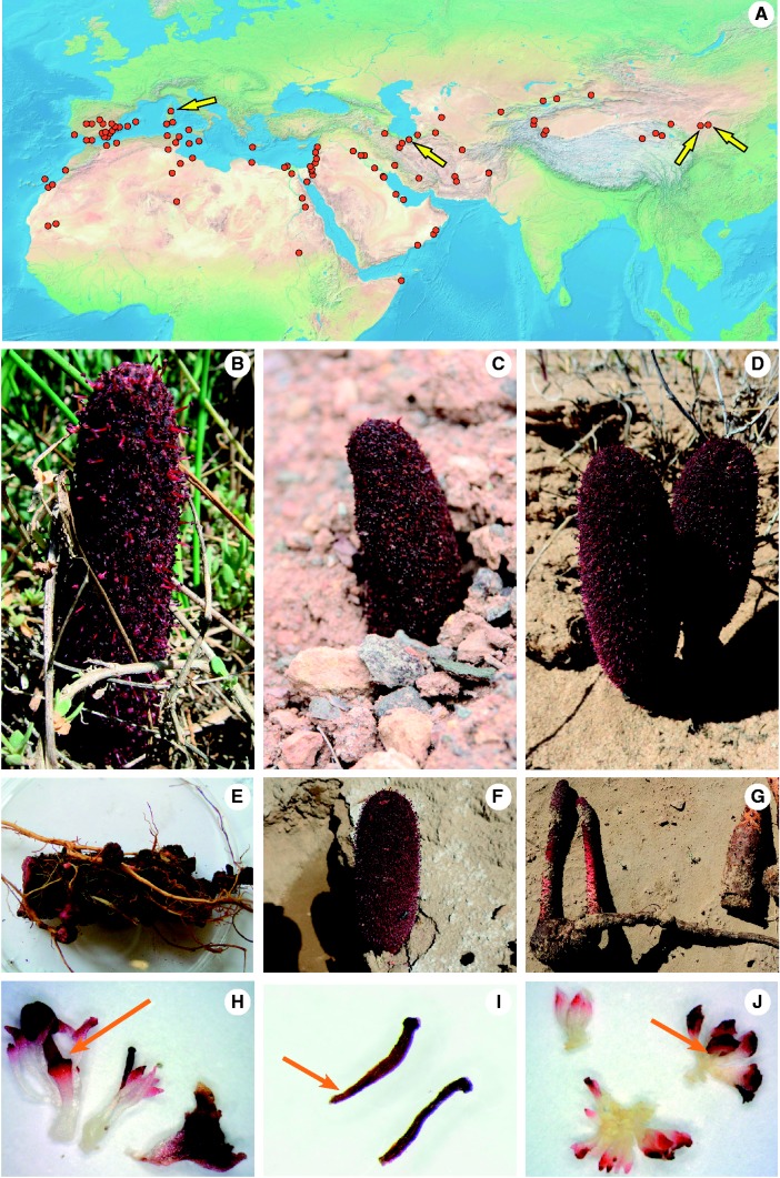
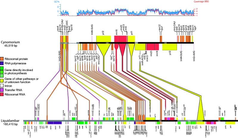
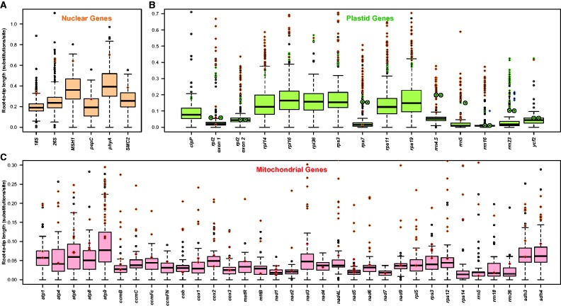
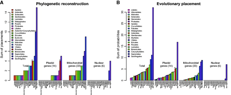
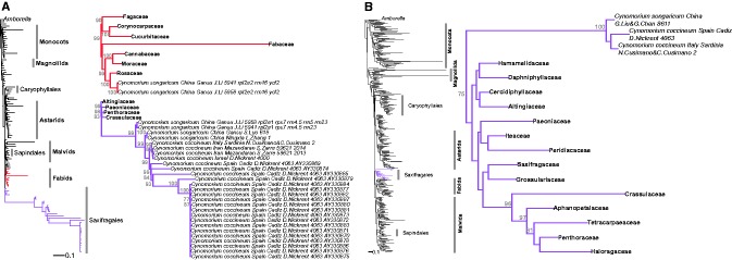
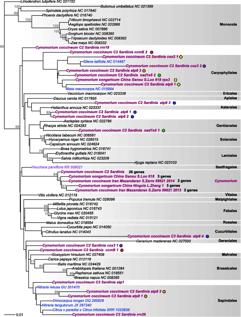
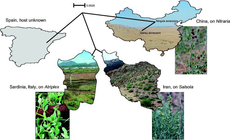
Cynomoriaceae, one of the last unplaced families of flowering plants, comprise one or two species or subspecies of root parasites that occur from the Mediterranean to the Gobi Desert. Using Illumina sequencing, we assembled the mitochondrial and plastid genomes as well as some nuclear genes of a Cynomorium specimen from Italy. Selected genes were also obtained by Sanger sequencing from individuals collected in China and Iran, resulting in matrices of 33 mitochondrial, 6 nuclear, and 14 plastid genes and rDNAs enlarged to include a representative angiosperm taxon sampling based on data available in GenBank. We also compiled a new geographic map to discern possible discontinuities in the parasites' occurrence. Cynomorium has large genomes of 13.70-13.61 (Italy) to 13.95-13.76 pg (China). Its mitochondrial genome consists of up to 49 circular subgenomes and has an overall gene content similar to that of photosynthetic angiosperms, while its plastome retains only 27 of the normally 116 genes. Nuclear, plastid and mitochondrial phylogenies place Cynomoriaceae in Saxifragales, and we found evidence for several horizontal gene transfers from different hosts, as well as intracellular gene transfers.

Keywords: Cynomorium; Mediterranean-Irano-Turanian; chondriome; horizontal gene transfer; parasitic plants; plastome.


Figures

Fig. 1.—
Fig. 1.—
Distribution and habit of Cynomorium . (A) Distribution range of Cynomorium obtained from the map in Hansen (1986) , relevant floras, and 203 GPS coordinates retrieved from the Global Biodiversity Information Facility (GBIF) portal in August 2015. Arrows indicate our own collections (details in table 1). Background map from Naturalearthdata.com. (B–J) Cynomorium plants from Italy (B, E, H), N. Cusimano and C. Cusimano 2, Iran (C and I) Zarre 59621 , and China (D) S.X. Luo 2014 from Tengger Desert; (F, G, J) G. Sun 1, from Gansu. Photos B to G by the respective collectors, photos H to J by N. Cusimano. (B–D) Plants in situ. (E) Part of a rhizome with young inflorescences and connected to the host roots ( Atriplex portulacoides ). (F) A fly ( Musca spec.) visiting Cynomorium in the Tengger Desert. (G) Chinese plant connected to the host root ( Nitraria tangutorum ). (H) Young male and female flowers and a bract, showing a red stamen basis (arrow). (I) Stamen from herbarium material. (J) Male flowers showing white stamen bases (arrow). The color of the stamen basis has been used to differentiate C. coccineum subsp. songaricum from Cynomorium coccineum subsp. coccineum .
Fig. 2.—
Fig. 2.—
Gene losses and rearrangements in the plastome of Cynomorium compared to that of Liquidambar formosana (GenBank accession KC588388). Colored lines link homologous genes between the two plastomes. The LSC of Cynomorium is divided in two parts (linked by the dotted line) to help visualization. Maps were drawn using OGDraw ( Lohse et al. 2013 ). GC content and coverage of the Cynomorium plastome are depicted in blue and red, respectively. (The pattern is the same when using less stringent mapping parameters; Results.).
Fig. 3.—
Fig. 3.—
Root to tip branch lengths of Cynomorium and other angiosperms in substitutions per site. Boxplots and open circles summarize the branch length distribution of: (A) nuclear, (B) plastid, and (C) mitochondrial genes of photosynthetic plants obtained from the constrained phylogenies in supplementary figures S3–S5 , Supplementary Material online. Black line: median; boxes: upper and lower quartile, including 50% of the data; whiskers: minimum and maximum of the data, provided that their length does not exceed 1.5× the interquartile range; open dots: outliers. Colored diamonds and circles represent, respectively, the branch length of the genes and of their copies found in other genomic compartments of Cynomorium : orange: gene copy located in the nuclear genome; green: gene copy located in the plastid genome; red: gene copy located in the mitochondrial genome; blue: gene copy amplified by Garcia et al. (2004) ; circles: Cynomorium gene copies amplified by Zhang et al. (2009) ; diamonds: all other Cynomorium sequences.
Fig. 4.—
Fig. 4.—
Bar plots summarizing the analyses of single gene matrices when the rest of the topology was constrained to fit the currently accepted angiosperm relationships ( Angiosperm Phylogeny Group 2016 ). For simplicity, internally transferred gene copies were not included but their placements can be seen in supplementary figures S4–S6 , Supplementary Material online. (A) Results of ML tree searches. For each clade, the number of genes that placed Cynomorium sequence(s) in the respective clade is shown (details in supplementary figs. S4–S6 and table S4 , Supplementary Material online). (B) Results of evolutionary placement analyses (Materials and Methods). For each clade, the sum of the probabilities across all genes that placed Cynomorium in the respective clade is shown (details in supplementary fig. S7 , Supplementary Material online). For both analyses, results are shown overall and by genomic compartment. The asterisks indicate placements resulting exclusively from the sequences of Zhang et al. (2009) . Numbers of markers for each compartment are indicated in brackets.
Fig. 5.—
Fig. 5.—
Phylogenetic trees obtained from ML analyses of the concatenated plastid (A) and nuclear (B) genes. Major angiosperm taxa are labeled following Angiosperm Phylogeny Group (2016) , with the orders including Cynomorium in purple (Saxifragales) and red (Fabids), and shown in more detail on the right.
Fig. 6.—
Fig. 6.—
Phylogenetic tree obtained from ML analyses of the concatenated mitochondrial gene matrix. The genus label Cynomorium on the right refers to the placement of 26 genes from the Italian plant. The colored dots mark multiple copies of the respective gene; the two yellow diamonds mark a gene acquired by HGT, of which one native copy was included in the concatenated matrix; the single yellow square marks the rps3 of a Chinese accession (see text).
Fig. 7.—
Fig. 7.—
Neighbor net obtained from variable plastid regions of Cynomorium from Spain ( Nickrent 4063 ), Italy (N. Cusimano and C. Cusimano 2), Iran ( S. Zarre 59621 ), and China (L. Zhang 1 and S.X. Luo 618). The photos show habitats and hosts of Cynomorium at our collecting sites. The host of the Spanish sample is unknown. Photos by N. Cusimano (Italy and Atriplex ), S.X. Luo (China and Nitraria ), and A. Gröger (Iran and Salsola ).

References

    1. Angiosperm Phylogeny Group . 2009. . An update of the Angiosperm Phylogeny Group classification for the orders and families of flowering plants: APG III . Bot J Linn Soc . 161 : 105 – 121 .
    1. Angiosperm Phylogeny Group . 2016. . An update of the Angiosperm Phylogeny Group classification for the orders and families of flowering plants: APG IV . Bot J Linn Soc . 181 : 1 – 20 .
    1. Barkman TJ , et al. . 2007. . Mitochondrial DNA suggests at least 11 origins of parasitism in angiosperms and reveals genomic chimerism in parasitic plants . BMC Evol Biol. 7 : 248.. - PMC - PubMed
    1. Bellot S, Renner SS. 2014. . Exploring new dating approaches for parasites: the worldwide Apodanthaceae (Cucurbitales) as an example . Mol Phylogenet Evol. 80 : 1 – 10 . - PubMed
    1. Bellot S, Renner SS. 2016. . The plastomes of two species in the endoparasite genus Pilostyles (Apodanthaceae) each retain just five or six possibly functional genes . Genome Biol Evol. 8 : 189 – 201 . - PMC - PubMed

Publication types

LinkOut - more resources