. 2016 Jun 29;36(26):6857-71.
doi: 10.1523/JNEUROSCI.4582-15.2016.

Muscle IL1β Drives Ischemic Myalgia via ASIC3-Mediated Sensory Neuron Sensitization

Affiliations

Muscle IL1β Drives Ischemic Myalgia via ASIC3-Mediated Sensory Neuron Sensitization

Jessica L Ross et al. J Neurosci. .

Abstract

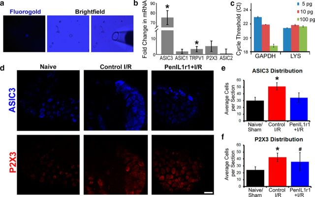
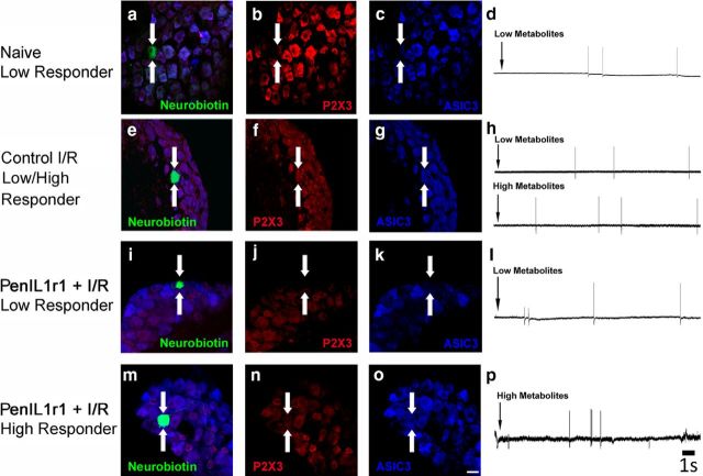
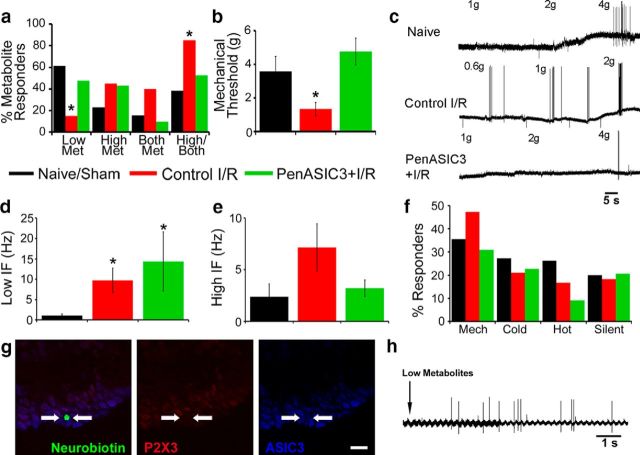
Musculoskeletal pain is a significantly common clinical complaint. Although it is known that muscles are quite sensitive to alterations in blood flow/oxygenation and a number of muscle pain disorders are based in problems of peripheral perfusion, the mechanisms by which ischemic-like conditions generate myalgia remain unclear. We found, using a multidisciplinary experimental approach, that ischemia and reperfusion injury (I/R) in male Swiss Webster mice altered ongoing and evoked pain-related behaviors in addition to activity levels through enhanced muscle interleukin-1 beta (IL1β)/IL1 receptor signaling to group III/IV muscle afferents. Peripheral sensitization depended on acid-sensing ion channels (ASICs) because treatment of sensory afferents in vitro with IL1β-upregulated ASIC3 in single cells, and nerve-specific knock-down of ASIC3 recapitulated the results of inhibiting the enhanced IL1β/IL1r1 signaling after I/R, which was also found to regulate afferent sensitization and pain-related behaviors. This suggests that targeting muscle IL1β signaling may be a potential analgesic therapy for ischemic myalgia.

Significance statement: Here, we have described a novel pathway whereby increased inflammation within the muscle tissue during ischemia/reperfusion injury sensitizes group III and IV muscle afferents via upregulation of acid-sensing ion channel 3 (ASIC3), leading not only to alterations in mechanical and chemical responsiveness in individual afferents, but also to pain-related behavioral changes. Furthermore, these I/R-induced changes can be prevented using an afferent-specific siRNA knock-down strategy targeting either ASIC3 or the upstream mediator of its expression, interleukin 1 receptor 1. Therefore, this knowledge may contribute to the development of alternative therapeutics for muscle pain and may be especially relevant to pain caused by issues of peripheral circulation, which is commonly observed in disorders such as complex regional pain syndrome, sickle cell anemia, or fibromyalgia.

Keywords: ASICs; behavior; cytokines; electrophysiology; siRNAs; signaling.


Figures

Figure 1.
Figure 1.
Increased IL1β signaling at the IL1r1 receptor regulates myalgia-related behaviors after I/R injury. Increased muscle IL1β protein expression 1 d after I/R (a, b) is localized to macrophages and neutrophils (arrows) present in injured muscles (c) as visualized with hematoxylin and eosin stain (left) or IL1β with hematoxylin only (right). n = 2. Expression of IL1r1 also increases in the affected DRGs (d, e), but nerve-specific siRNA knock-down of IL1r1 (PenIL1r1 + I/R) prevents this I/R-induced upregulation (n = 3). Knock-down of the receptor likely affected all neuronal cell types because IL1r1 was immunocytochemically localized (n = 3) to most DRG neurons (f). g, ATF3 (red) was used to mark injured neurons (arrows) in the affected C8 or T1 DRGs 1 d after either a median and ulnar nerve crush or an injection of nontargeting, control siRNAs (PenCON) into the median and ulnar nerves (n = 2–3 per condition). Crush injury readily induces ATF3 expression in DRGs, whereas PenCON injection does not. Behavioral deficits caused by I/R are also attenuated with IL1r1 knock-down (n ≥ 8 per injury condition per task). h, Spontaneous paw-guarding behaviors increase significantly 1 d after injury in the control I/R group compared with PenIL1r1 + I/R and naive/sham animals. Furthermore, control I/R animals showed a significant improvement within their group on D3 compared with D1 function (p < 0.001), whereas the other groups showed no significant guarding behaviors over time (p ≥ 0.101). Both paw withdrawal threshold (i) and grip strength (j) decreased significantly 1 d after I/R, but these effects were attenuated by IL1r1 knock-down. On D3, the effects of I/R alone had resolved in both tests. k, Voluntary activity is significantly decreased within 1 d after I/R, but is no different from PenIL1r1 + I/R or naive/sham animals by D3. Average velocity is also not different between groups on any testing day (l). Data are represented as mean ± SEM and were analyzed via one-way ANOVA with Tukey's post hoc or two-way RM ANOVA with Holm–Sidak post hoc tests. *p < 0.05 compared with naive/sham.
Figure 2.
Figure 2.
Nerve-specific siRNA knock-down of IL1r1 alters I/R-induced changes in group III and IV muscle afferent phenotypes and response properties as assessed by ex vivo recording. a, Compared with naive muscle afferents, I/R decreases the total number of cells responding to low (innocuous) metabolite solutions and increases the number of cells responsive to either high (noxious) metabolites or both of these solutions; however, afferent phenotypes recorded in the PenIL1r1 + I/R condition were no different from naïve. *p < 0.05, χ2 analysis. n ≥ 45 characterized cells per condition. b, IL1r1 knock-down also prevents the I/R-associated decrease in mechanical threshold. c, Examples of responses to mechanical deformation of the muscles in naive/sham, control I/R, and PenIL1r1 + I/R mice. d, Although I/R caused increased firing to low metabolites, nerve-specific inhibition of IL1r1 did not restore mean peak instantaneous firing frequencies to this metabolite solution back to naive levels. e, Instantaneous frequencies to high metabolites were also not different in any group. f, I/R did not alter total numbers of thermally sensitive, mechanically responsive, or silent fibers and siRNA knock-down of IL1r1 also did not affect these phenotypes. Data are represented as mean ± SEM and were analyzed via Kruskal–Wallis one-way ANOVA on ranks with Dunn's test. *p < 0.05 compared with naive.
Figure 3.
Figure 3.
ASIC3 upregulation after I/R is linked to increased IL1β and IL1r1 expression. a, FG-labeled primary afferents from DRG cultures (n = 3–6) for single-cell qPCR. b, Cells incubated in IL1β-enriched medium showed significant upregulation of ASIC3 and TRPV1, but not P2X3, ASIC1, or ASIC2, compared with untreated cells (n = 10–12 cells per treatment). c, Verification of LYS internal control for use in single-cell real-time PCR. We performed our single-cell qPCR analysis from whole DRG RNA samples diluted to varying starting concentrations (5, 10, and 100 pg) containing the same amount of spike RNA (LYS: 1000 copies). Analysis of these postamplification samples shows that, even when varying the total starting RNA concentration (and thus the Ct values for GAPDH), we still obtain a consistent level of the spike RNA. d, Representative immunohistochemical staining of ASIC3 or P2X3 protein in whole DRGs from naive, control I/R, and IL1r1 conditions are shown. ASIC3-immunopositive cells are significantly increased in the control I/R condition over both the naive and PenIL1r1 + I/R conditions, which did not differ (e). Increased P2X3 after I/R was not affected by IL1r1 knock-down (f). Data are represented as mean ± SEM and were analyzed via one-way ANOVA with Tukey's post hoc test. *p < 0.05 compared with naive or untreated; #p > 0.05 compared with naive and control I/R. Scale bar for all images, 60 μm.
Figure 4.
Figure 4.
ASIC3 expression in naive animals is present in metabo-nociceptors. a, Examples of a Lucifer yellow-labeled muscle afferent recovered in vitro after characterization via ex vivo recording (n = 6). b, This “high” responder was found to be ASIC3-positive at the mRNA level. Presence of GAPDH verified appropriately amplified cDNAs in the single cell. c, An intracellularly stained (neurobiotin, green) and physiologically characterized muscle afferent (arrows) from uninjured tissue that was responsive to high metabolites (d, e) was also immunopositive for both ASIC3 (blue) and P2X3 (red). Scale bar for all images, 60 μm.
Figure 5.
Figure 5.
Neurochemical identification of single metabolite-responsive muscle afferents recovered from ex vivo recordings in each condition. A neurobiotin-labeled (green, arrows) naive muscle afferent (a) that was responsive to low, but not high (d), metabolites was found to be immunonegative for both P2X3 (red) and ASIC3 (blue; b, c). After 1 d I/R, a chemosensitive muscle afferent (e) sensitive to both metabolite solutions (h) was immunopositive for P2X3 and ASIC3 (f, g). Characterized and labeled metabolite-sensitive afferents from the PenIL1r1 + I/R conditions showed differing neurochemical phenotypes based on their metabolite selectivity (ip). A neuron responsive to low metabolites was found to be P2X3 and ASIC3 immunonegative (j, k), whereas a high responder from this condition was P2X3 and ASIC3 immunopositive (n, o). Scale bar for all images, 60 μm.
Figure 6.
Figure 6.
ASIC3 inhibition through siRNA-mediated knock-down attenuates I/R-induced alterations in voluntary activity and other pain-related behaviors. Total protein expression of ASIC3 is increased in the affected C7/C8/T1 DRGs after I/R (a), but channel upregulation is blocked by nerve-specific siRNA knock-down of either IL1r1 or ASIC3 (n = 3). b, PenASIC3 + I/R animals were no different from naive/sham animals in spontaneous pain behaviors measured by the paw-guarding assay on D1 or D3 and they did not significantly change from BL measurements at either time point. c, Paw withdrawal thresholds in the PenASIC3 + I/R group also did not change from BL levels; however, the I/R-induced grip strength deficit was not completely prevented by ASIC3 knock-down (d) because PenASIC3 + I/R animals did not differ significantly from either control I/R or naive/sham animals at D1, even though there appeared to be significant improvement between D1 and D3 in the PenASIC3 + I/R condition. On D1, PenASIC3 + I/R animals ran significantly greater distances (e) than control I/R mice. They also ran at higher velocities (f) than either the naive/sham or control I/R groups. This velocity difference also performed to D3 compared with the control I/R animals, but not to any other group. Data are represented as mean ± SEM and were analyzed via one-way ANOVA with Tukey's post hoc or two-way RM ANOVA with Holm–Sidak post hoc tests. n ≤ 8 for each behavioral task. *p < 0.05 compared with all other conditions; #p < 0.05 compared with naive/sham.
Figure 7.
Figure 7.
Inhibition of post-I/R ASIC3 upregulation prevents injury-induced alterations in muscle afferents. a, Similar to siRNA-mediated IL1r1 knock-down, group III and IV primary muscle afferent phenotypes in the PenASIC3 + I/R condition are no different from the naive condition as assessed with ex vivo recording. b, ASIC3 knock-down also prevents the I/R-associated decrease in mechanical threshold. c, Examples of responses to mechanical deformation of the muscles in naive/sham, control I/R, and PenASIC3 + I/R mice. d, Although I/R caused increased firing to low metabolites, ASIC3-targeting siRNA injection did not restore mean peak instantaneous firing frequencies to this metabolite solution back to naive levels. e, Instantaneous frequency to high metabolites was also not different in any group. f, I/R did not alter total numbers of thermally sensitive, mechanically responsive, or silent fibers and siRNA knock-down of ASIC3 also did not affect these phenotypes. g, A neurobiotin-labeled (green, arrows) muscle afferent recovered from a PenASIC3 + I/R preparation that was responsive to low, but not high (h), metabolites was found to be immunonegative for both P2X3 (red) and ASIC3 (blue). Data are represented as mean ± SEM and were analyzed via χ2 analysis or Kruskal–Wallis one-way ANOVA on ranks with Dunn's test. *p < 0.05 compared with naive. Scale bar for all images, 60 μm.
Figure 8.
Figure 8.
I/R induces ASIC3 expression through activation/phosphorylation of JNK. a, Representative immunohistochemical staining of ASIC3 (blue) and p-JNK (red) protein in whole DRGs. Several p-JNK/ASIC3 double-labeled cells (arrows) were found in both conditions, as well as p-JNK single-labeled (arrowheads) and ASIC3 single-labeled (dashed arrowheads) cells. Shortly after I/R (3 h), DRG expression of p-JNK significantly increases (b), as does p-JNK/ASIC3 coexpression (c) relative to naive conditions. d, Representative immunohistochemical staining of p-JNK (red) protein in primary afferents retrogradely labeled from the forepaw muscles with FG (blue, arrows) and cultured in untreated medium or medium enriched with IL1β. IL1β treatment increases the numbers of p-JNK-positive cells in vitro (e) and increases the expression of ASIC3 mRNA (f) in individual muscle afferents. However, treatment of IL1β-exposed cells with a JNK inhibitor (SP600125), but not an NFκB inhibitor (CAPE), completely prevents ASIC3 upregulation in single cells. g, Schematic of AP1/Jun transcription factor binding sites on ASIC3 promoter region. Data are represented as mean ± SEM and were analyzed via one-way ANOVA with Holm–Sidak test. *p < 0.05 compared with other conditions. Scale bar, 60 μm.

Similar articles

Cited by

References

    1. Alam M, Smirk FH. Observations in man upon a blood pressure raising reflex arising from the voluntary muscles. J Physiol. 1937;89:372–383. doi: 10.1113/jphysiol.1937.sp003485. - DOI - PMC - PubMed
    1. Ali FN, Carman TL. Medical management for chronic atherosclerotic peripheral arterial disease. Drugs. 2012;72:2073–2085. doi: 10.2165/11640810-000000000-00000. - DOI - PubMed
    1. Alvarez P, Green PG, Levine JD. Role for monocyte chemoattractant protein-1 in the induction of chronic muscle pain in the rat. Pain. 2014;155:1161–1167. doi: 10.1016/j.pain.2014.03.004. - DOI - PMC - PubMed
    1. Benson CJ, McCleskey EW. ASICs function as lactic acid sensors during cardiac ischemia. In: Wang DH, editor. Molecular sensors for cardiovascular homeostasis. New York: Springer; 2007. pp. 32–50.
    1. Benson CJ, Eckert SP, McCleskey EW. Acid-evoked currents in cardiac sensory neurons: a possible mediator of myocardial ischemic sensation. Circ Res. 1999;84:921–928. doi: 10.1161/01.RES.84.8.921. - DOI - PubMed

Publication types

MeSH terms

LinkOut - more resources