

Implications of Lateral Cerebellum in Proactive Control of Saccades
- PMID: 27358462
- PMCID: PMC6604892
- DOI: 10.1523/JNEUROSCI.0733-16.2016
Implications of Lateral Cerebellum in Proactive Control of Saccades
Abstract
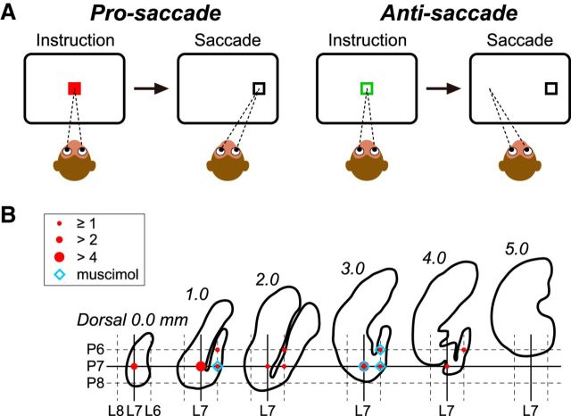
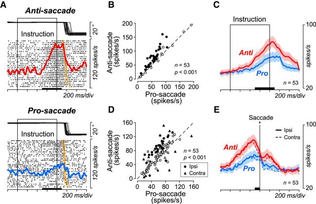
Although several lines of evidence establish the involvement of the medial and vestibular parts of the cerebellum in the adaptive control of eye movements, the role of the lateral hemisphere of the cerebellum in eye movements remains unclear. Ascending projections from the lateral cerebellum to the frontal and parietal association cortices via the thalamus are consistent with a role of these pathways in higher-order oculomotor control. In support of this, previous functional imaging studies and recent analyses in subjects with cerebellar lesions have indicated a role for the lateral cerebellum in volitional eye movements such as anti-saccades. To elucidate the underlying mechanisms, we recorded from single neurons in the dentate nucleus of the cerebellum in monkeys performing anti-saccade/pro-saccade tasks. We found that neurons in the posterior part of the dentate nucleus showed higher firing rates during the preparation of anti-saccades compared with pro-saccades. When the animals made erroneous saccades to the visual stimuli in the anti-saccade trials, the firing rate during the preparatory period decreased. Furthermore, local inactivation of the recording sites with muscimol moderately increased the proportion of error trials, while successful anti-saccades were more variable and often had shorter latency during inactivation. Thus, our results show that neuronal activity in the cerebellar dentate nucleus causally regulates anti-saccade performance. Neuronal signals from the lateral cerebellum to the frontal cortex might modulate the proactive control signals in the corticobasal ganglia circuitry that inhibit early reactive responses and possibly optimize the speed and accuracy of anti-saccades.
Significance statement: Although the lateral cerebellum is interconnected with the cortical eye fields via the thalamus and the pons, its role in eye movements remains unclear. We found that neurons in the caudal part of the lateral (dentate) nucleus of the cerebellum showed the increased firing rate during the preparation of anti-saccades. Inactivation of the recording sites modestly elevated the rate of erroneous saccades to the visual stimuli in the anti-saccade trials, while successful anti-saccades during inactivation tended to have a shorter latency. Our data indicate that neuronal signals in the lateral cerebellum may proactively regulate anti-saccade generation through the pathways to the frontal cortex, and may inhibit early reactive responses and regulate the accuracy of anti-saccades.
Keywords: anti-saccade; cerebellum; dentate nucleus; inactivation; primate; single neurons.
Copyright © 2016 the authors 0270-6474/16/367066-09$15.00/0.
Figures


Similar articles
-
Inactivation of macaque lateral intraparietal area delays initiation of the second saccade predominantly from contralesional eye positions in a double-saccade task.Exp Brain Res. 2001 Mar;137(1):45-57. doi: 10.1007/s002210000546. Exp Brain Res. 2001. PMID: 11310171
-
Cerebellar Roles in Self-Timing for Sub- and Supra-Second Intervals.J Neurosci. 2017 Mar 29;37(13):3511-3522. doi: 10.1523/JNEUROSCI.2221-16.2017. Epub 2017 Feb 27. J Neurosci. 2017. PMID: 28242799 Free PMC article.
-
Effects of reversible inactivation of the primate mesencephalic reticular formation. I. Hypermetric goal-directed saccades.J Neurophysiol. 2000 Apr;83(4):2260-84. doi: 10.1152/jn.2000.83.4.2260. J Neurophysiol. 2000. PMID: 10758133
-
Cerebellar control of saccadic eye movements: its neural mechanisms and pathways.Jpn J Physiol. 1991;41(3):351-68. doi: 10.2170/jjphysiol.41.351. Jpn J Physiol. 1991. PMID: 1960885 Review.
-
[Functional analysis of the thalamocortical pathways in eye movements].Brain Nerve. 2011 Aug;63(8):871-7. Brain Nerve. 2011. PMID: 21817178 Review. Japanese.
Cited by
-
Consensus Paper: Neurophysiological Assessments of Ataxias in Daily Practice.Cerebellum. 2018 Oct;17(5):628-653. doi: 10.1007/s12311-018-0937-2. Cerebellum. 2018. PMID: 29656311 Review.
-
Modular output circuits of the fastigial nucleus for diverse motor and nonmotor functions of the cerebellar vermis.Elife. 2020 Jul 8;9:e58613. doi: 10.7554/eLife.58613. Elife. 2020. PMID: 32639229 Free PMC article.
-
Different contributions of preparatory activity in the basal ganglia and cerebellum for self-timing.Elife. 2018 Jul 2;7:e35676. doi: 10.7554/eLife.35676. Elife. 2018. PMID: 29963985 Free PMC article.
-
Long-Term Predictive and Feedback Encoding of Motor Signals in the Simple Spike Discharge of Purkinje Cells.eNeuro. 2017 Apr 11;4(2):ENEURO.0036-17.2017. doi: 10.1523/ENEURO.0036-17.2017. eCollection 2017 Mar-Apr. eNeuro. 2017. PMID: 28413823 Free PMC article.
-
Maturation of Temporal Saccade Prediction from Childhood to Adulthood: Predictive Saccades, Reduced Pupil Size, and Blink Synchronization.J Neurosci. 2022 Jan 5;42(1):69-80. doi: 10.1523/JNEUROSCI.0837-21.2021. Epub 2021 Nov 10. J Neurosci. 2022. PMID: 34759032 Free PMC article.
References
-
- Arikan R, Blake NM, Erinjeri JP, Woolsey TA, Giraud L, Highstein SM. A method to measure the effective spread of focally injected muscimol into the central nervous system with electrophysiology and light microscopy. J Neurosci Methods. 2002;118:51–57. - PubMed
Publication types
MeSH terms
Substances
LinkOut - more resources
Full Text Sources
Other Literature Sources