. 2017 Feb 1;23(3):677-686.
doi: 10.1158/1078-0432.CCR-16-0558. Epub 2016 Jun 29.

HER3 Targeting Sensitizes HNSCC to Cetuximab by Reducing HER3 Activity and HER2/HER3 Dimerization: Evidence from Cell Line and Patient-Derived Xenograft Models

Affiliations

HER3 Targeting Sensitizes HNSCC to Cetuximab by Reducing HER3 Activity and HER2/HER3 Dimerization: Evidence from Cell Line and Patient-Derived Xenograft Models

Dongsheng Wang et al. Clin Cancer Res. .

Abstract

Purpose: Our previous work suggested that HER3 inhibition sensitizes head and neck squamous cell carcinoma (HNSCC) to EGFR inhibition with cetuximab. This study aimed to define the role of HER3 in cetuximab resistance and the antitumor mechanisms of EGFR/HER3 dual targeting in HNSCC.

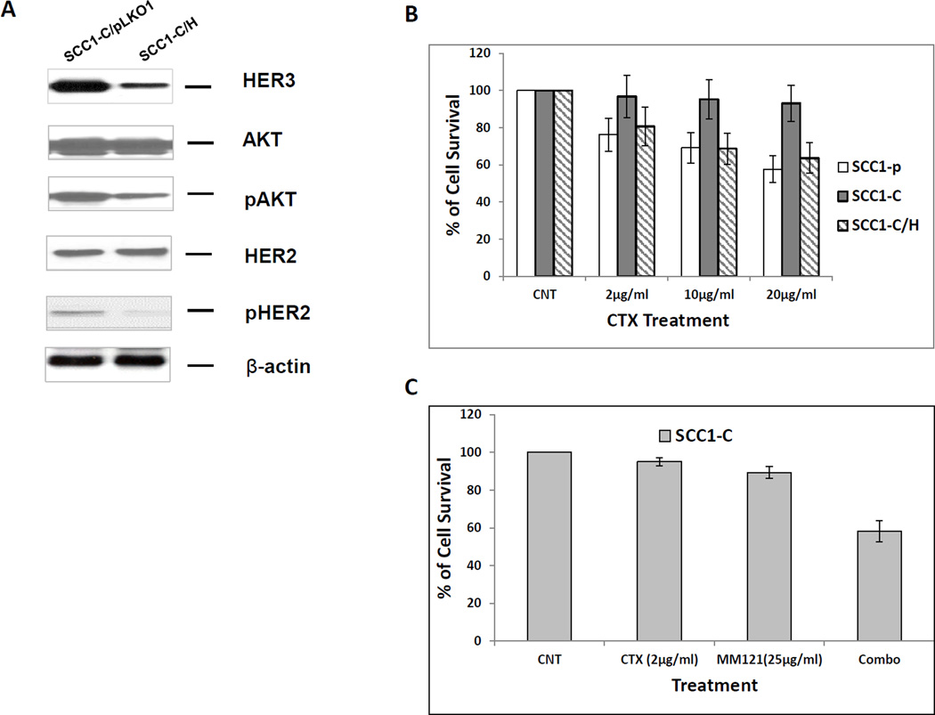
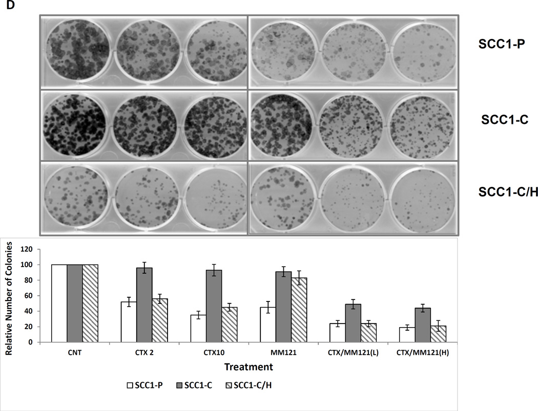
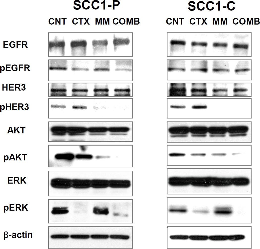
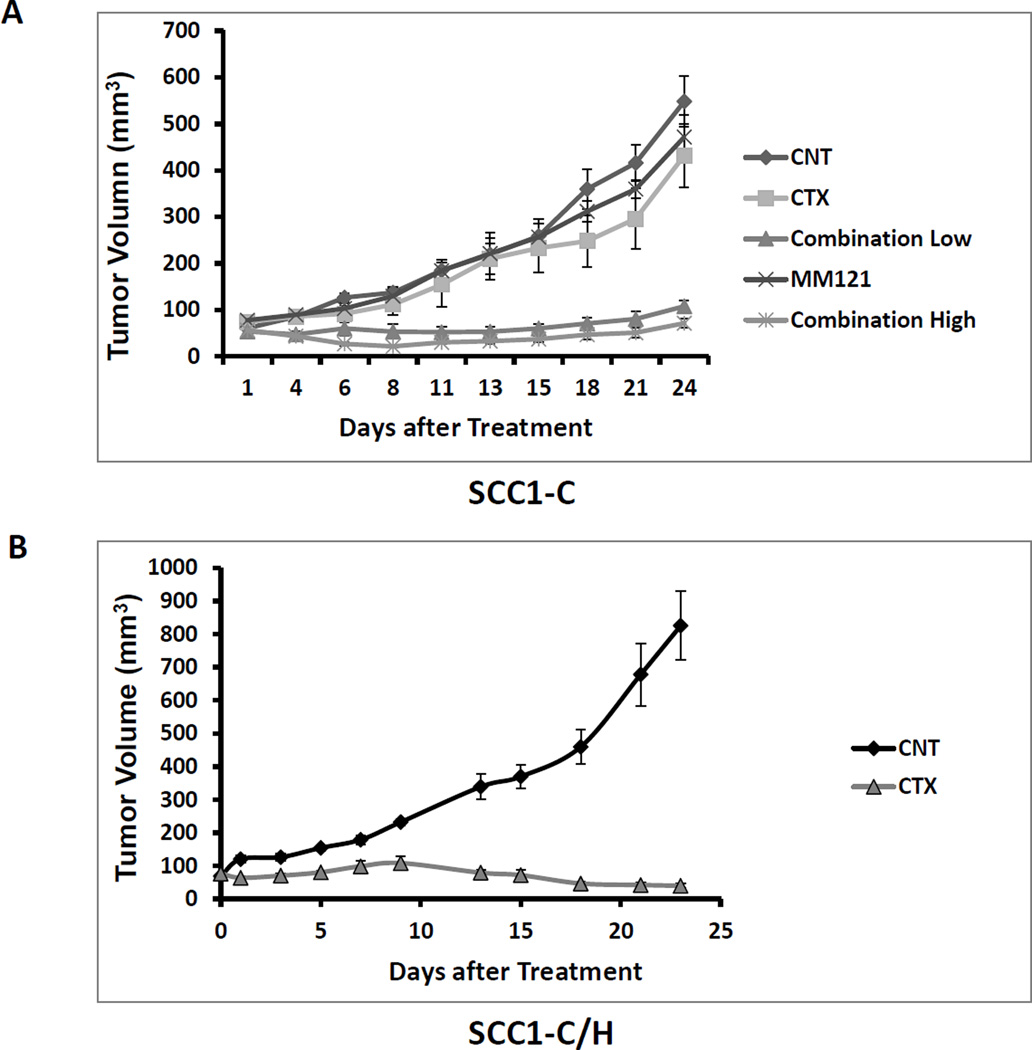
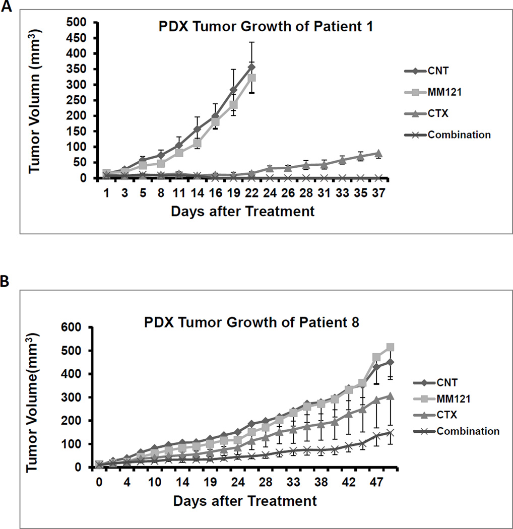
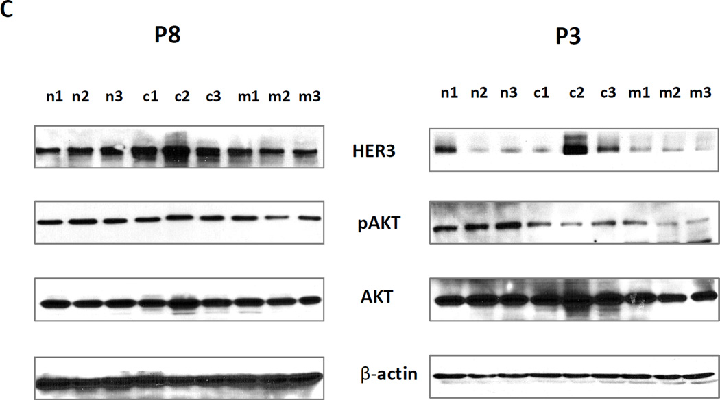
Experimental design: We treated cetuximab-resistant HNSCC UMSCC1-C and parental UMSCC1-P cell lines with anti-EGFR antibody cetuximab, anti-HER3 antibody MM-121, and their combination. We assessed activities of HER2, HER3, and downstream signaling pathways by Western blotting and cell growth by sulforhodamine B (SRB) and colony formation assays. HER3-specific shRNA was used to confirm the role of HER3 in cetuximab response. The combined efficacy and alterations in biomarkers were evaluated in UMSCC1-C xenograft and patient-derived xenograft (PDX) models.

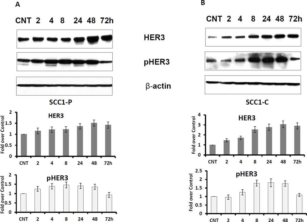
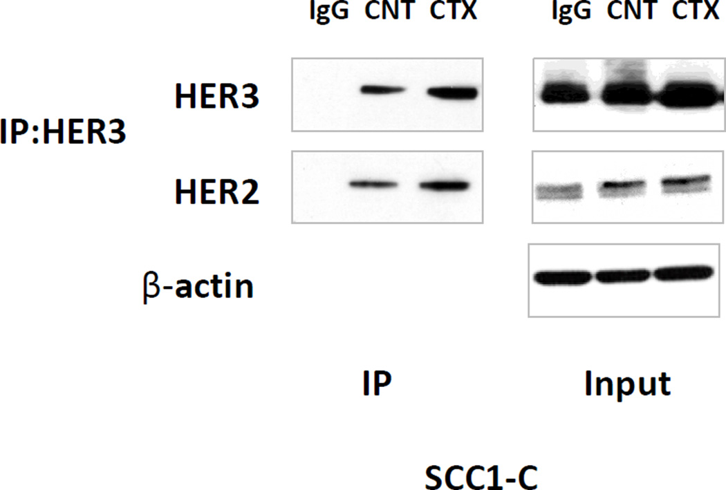
Results: Cetuximab treatment induced HER3 activation and HER2/HER3 dimerization in HNSCC cell lines. Combined treatment with cetuximab and MM-121 blocked EGFR and HER3 activities and inhibited the PI3K/AKT and ERK signaling pathways and HNSCC cell growth more effectively than each antibody alone. HER3 knockdown reduced HER2 activation and resensitized cells to cetuximab. Cetuximab-resistant xenografts and PDX models revealed greater efficacy of dual EGFR and HER3 inhibition compared with single antibodies. In PDX tissue samples, cetuximab induced HER3 expression and MM-121 reduced AKT activity.

Conclusions: Clinically relevant PDX models demonstrate that dual targeting of EGFR and HER3 is superior to EGFR targeting alone in HNSCC. Our study illustrates the upregulation of HER3 by cetuximab as one mechanism underlying resistance to EGFR inhibition in HNSCC, supporting further clinical investigations using multiple targeting strategies in patients who have failed cetuximab-based therapy. Clin Cancer Res; 23(3); 677-86. ©2016 AACR.


Conflict of interest statement

of Potential Conflicts of Interest No potential conflicts of interest to disclose.

Figures

Fig 1
Fig 1. Cetuximab induces HER3 expression and activation in HNSCC cell lines
Cetuximab sensitive UMSCC1-P cells (A) and counterpart resistant UMSCC1-C cells (B) were cultured in medium with 5% FBS. The cells were treated with 2µg/ml cetuximab for the indicated times. CNT indicates the control sample. Both HER3 expression and activation (pHER3) levels were elevated by cetuximab treatment in a time dependent manner (Figure represents 1 of 3 experiments). The average fold increase was determined from 3 individual experiments with standard deviations.
Fig 2
Fig 2. HER2/HER3 dimerization is increased upon cetuximab treatment
UMSCC1-C cells were exposed to 2µg/ml cetuximab for 24 hr. HER3 was immunoprecipitated from the cell lysate with anti-HER3 antibody. The immunoprecipitate was fractionated on SDS-PAGE followed by immunoblotting with anti-HER2 and HER3 antibodies. As the image shows, cetuximab treatment increased HER3 expression in UMSCC1-C cells, at the same time HER2 and HER3 association was increased as more HER2 was detected by immunoblot in cetuximab treated cells compared with control cells and IgG control. β-actin was used as loading control (figure represents 1 of 3 experiments).
Fig 3
Fig 3. Inhibition of HER3 re-sensitizes resistant UMSCC1-C cell line to cetuximab
(A) HER was knocked down in UMSCC1-C/H cells. HER2 and AKT activities were also reduced, as demonstrated by a decrease in both pHER2 and pAKT levels. (B) SRB assay shows that cetuximab reduces the growth rate of sensitive parental UMSCC1-P cells at the indicated concentrations after treatment for 48 h. No growth inhibition was observed in UMSCC1-C cells. Knock down of HER3 by shRNA re-sensitized UMSCC1-C cells to cetuximab inhibition. (C) Combination of cetuximab (2µg/ml) and MM-121 (25µg/ml)(combo) more potently inhibited UMSCC1-C growth in SRB assay. (D) In a colony formation assay, cetuximab inhibited colony formation of UMSCC1-P cells but not UMSCC1-C cells at the indicated concentration. When HER3 was knocked down by shRNA, cetuximab inhibition of colony formation was restored. Inhibition of HER3 by its antibody MM-121 (25 µg/ml) also resensitized UMSCC1-C to cetuximab treatment. (CTX2: cetuximab 2µg/ml, CTX10: cetuximab 10µg/ml, CTX/MM-121(L): combination of cetuximab 2µg/ml and MM-121, CTX/MM-121(H): combination of cetuximab 10µg/ml and MM-121). Image represents 3 individual experiments.
Fig 4
Fig 4. Combination of cetuximab and MM-121 inhibits both PI3K/AKT and ERK signaling pathways
UMSCC1-P (A) and UMSCC1-C (B) cells were treated with 2µg/mL cetuximab, 125µg/mL MM-121, and the combination, respectively. As shown in (A) and (B), after 48 hours of treatment, AKT and ERK activation was simultaneously ablated by the combination compared to single drugs and the control. The inhibition of pAKT was greater by MM-121 than cetuximab in both cell lines (figure represents 1 of 3 experiments).
Fig 5
Fig 5. Combination of cetuximab and MM-121 shows strong antitumor activity in HNSCC cetuximab resistant tumor xenograft animal model
(A) In xenograft models using cetuximab resistant UMSCC1-C cells, mice were randomly assigned to five treatment groups: PBS control, cetuximab (100µg/dose), MM-121 (300ug/dose), Comb.LD (cetuximab 100µg/dose/MM-121 300ug/dose), and Comb.HD (cetuximab 100µg/dose/MM-121 600ug/dose) and were treated twice a week through intraperitoneal injection. Consistent with our in vitro observations, the CM combination showed the greatest tumor growth inhibition in UMSCC1-C xenografts. Neither cetuximab nor MM-121 alone significantly reduced the tumor growth compared to PBS control. However, the groups treated with both high and low doses of CM combination showed significantly suppressed tumor growth as compared with those treated with PBS control, cetuximab and MM-121 alone. (p<0.001, n=6). (B) Mice carrying UMSCC1-C/H cell xenografts were randomly assigned to two treatment groups: PBS control, cetuximab (100µg/dose), Treatment with cetuximab significantly inhibited tumor growth (p<0.001, n=6).
Fig 6
Fig 6. Combination of anti-HER3 antibody MM-121 and cetuximab more potently inhibits tumor growth in HNSCC patient derived tumor xenografts than single treatment with only MM-121 or cetuximab alone
(A) In PDXs derived from Patient 1 tumor tissue, both cetuximab (100µg/dose) and the combination (cetuximab 100µg/dose and MM-121 300ug/dose) significantly inhibited tumor growth in nude mice (p<0.001 for both treatments). There was no significant difference between cetuximab alone and the combination. However, tumors treated with cetuximab alone grew back while no tumor relapse could be detected in the combination group. (B) In PDXs derived from Patient 8, only the combination treatment significantly inhibited tumor growth (p<0.004). Combination treatment was also more effective than either single agent (p<0.032 respectively). (C) As shown by immunoblotting, HER3 expression was induced in cetuximab treated PDX tumor samples, while pAKT was reduced by MM-121 treatment. No samples from the combination-treated PDXs were available for testing as the tumors were too small to isolate sufficient protein sample. n1–3: PDX tumor samples from 3 different PDXs in the control group. c1–3: three different tumor samples from the cetuximab treated group. m1–3: three different tumor samples from the MM-121 treated group. Image represents 3 repeated experiments.

References

    1. Ongkeko WM, Altuna X, Weisman RA, Wang-Rodriguez J. Expression of protein tyrosine kinases in head and neck squamous cell carcinomas. Am J Clin Pathol. 2005;124:71–76. - PubMed
    1. Bei R, Budillon A, Masuelli L, Cereda V, Vitolo D, et al. Frequent overexpression of multiple ErbB receptors by head and neck squamous cell carcinoma contrasts with rare antibody immunity in patients. J Pathol. 2004;204:317–325. - PubMed
    1. Vermorken JB, Mesia R, Rivera F, Remenar E, Kawecki A, et al. Platinum-based chemotherapy plus cetuximab in head and neck cancer. N Engl J Med. 2008;359:1116–1127. - PubMed
    1. Vermorken JB, Trigo J, Hitt R, Koralewski P, Diaz-Rubio E, et al. Open-label, uncontrolled, multicenter phase II study to evaluate the efficacy and toxicity of cetuximab as a single agent in patients with recurrent and/or metastatic squamous cell carcinoma of the head and neck who failed to respond to platinum-based therapy. J Clin Oncol. 2007;25:2171–2177. - PubMed
    1. Shaib W, Kono S, Saba N. Antiepidermal growth factor receptor therapy in squamous cell carcinoma of the head and neck. J Oncol. 2012;2012:521215. - PMC - PubMed

MeSH terms