. 2017 Jan 1;23(1):124-136.
doi: 10.1158/1078-0432.CCR-15-1535. Epub 2016 Jun 29.

Combination Therapy with Anti-PD-1, Anti-TIM-3, and Focal Radiation Results in Regression of Murine Gliomas

Affiliations

Combination Therapy with Anti-PD-1, Anti-TIM-3, and Focal Radiation Results in Regression of Murine Gliomas

Jennifer E Kim et al. Clin Cancer Res. .

Abstract

Purpose: Checkpoint molecules like programmed death-1 (PD-1) and T-cell immunoglobulin mucin-3 (TIM-3) are negative immune regulators that may be upregulated in the setting of glioblastoma multiforme. Combined PD-1 blockade and stereotactic radiosurgery (SRS) have been shown to improve antitumor immunity and produce long-term survivors in a murine glioma model. However, tumor-infiltrating lymphocytes (TIL) can express multiple checkpoints, and expression of ≥2 checkpoints corresponds to a more exhausted T-cell phenotype. We investigate TIM-3 expression in a glioma model and the antitumor efficacy of TIM-3 blockade alone and in combination with anti-PD-1 and SRS.

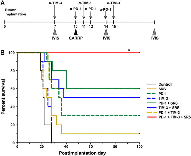
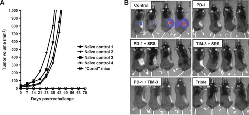
Experimental design: C57BL/6 mice were implanted with murine glioma cell line GL261-luc2 and randomized into 8 treatment arms: (i) control, (ii) SRS, (iii) anti-PD-1 antibody, (iv) anti-TIM-3 antibody, (v) anti-PD-1 + SRS, (vi) anti-TIM-3 + SRS, (vii) anti-PD-1 + anti-TIM-3, and (viii) anti-PD-1 + anti-TIM-3 + SRS. Survival and immune activation were assessed.

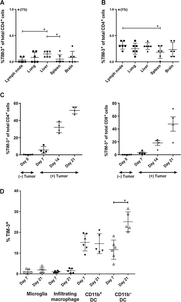
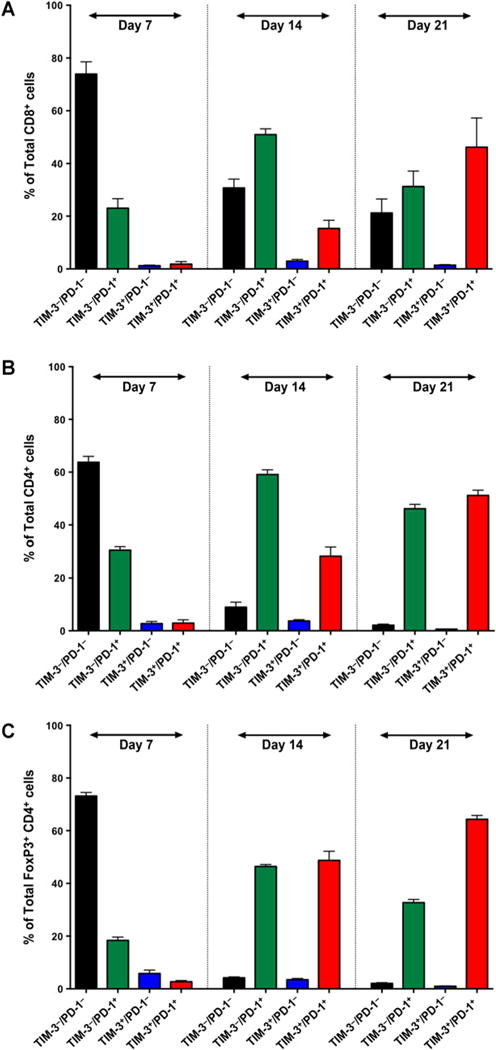
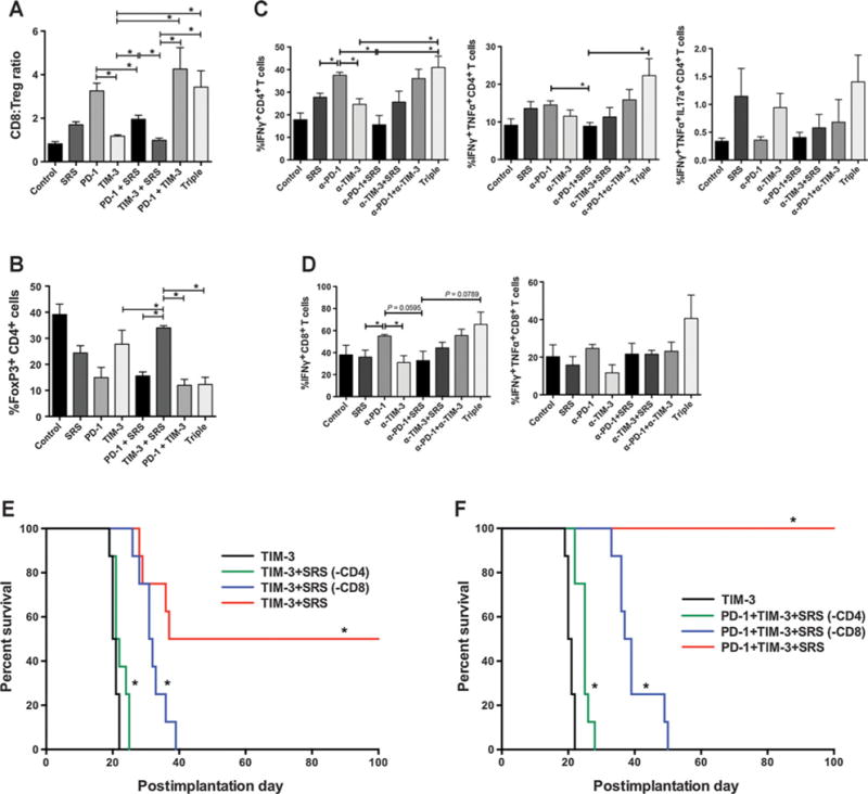
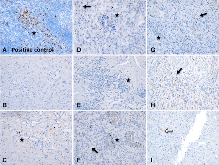
Results: Dual therapy with anti-TIM-3 antibody + SRS or anti-TIM-3 + anti-PD-1 improved survival compared with anti-TIM-3 antibody alone. Triple therapy resulted in 100% overall survival (P < 0.05), a significant improvement compared with other arms. Long-term survivors demonstrated increased immune cell infiltration and activity and immune memory. Finally, positive staining for TIM-3 was detected in 7 of 8 human GBM samples.

Conclusions: This is the first preclinical investigation on the effects of dual PD-1 and TIM-3 blockade with radiation. We also demonstrate the presence of TIM-3 in human glioblastoma multiforme and provide preclinical evidence for a novel treatment combination that can potentially result in long-term glioma survival and constitutes a novel immunotherapeutic strategy for the treatment of glioblastoma multiforme. Clin Cancer Res; 23(1); 124-36. ©2016 AACR.


Conflict of interest statement

Disclosure of Potential Conflicts of Interest

No potential conflicts of interest were disclosed by the other authors.

Figures

Figure 1
Figure 1
TIM-3 expression on peripheral and CNS lymphocytes. In naïve, non-tumor–bearing mice, less than 0.5% of all CD4+ (A) and CD8+ T cells (B) isolated from peripheral lymph nodes, lungs, livers, spleens, and brains had surface expression of TIM-3. C, Compared with naïve mice, tumor-bearing mice demonstrate a progressive increase in TIM-3 expression. From days 7, 14, and 21 postimplantation, the percentage of CD4+ T cells expressing TIM-3 rose from 5.65 ± 1.98% to 32 ± 3.16% to 51.75 ± 2.03%, respectively (P < 0.05). The percentage of CD8+ T cells rose from 3.05 ± 0.05% to 18.28 ± 3.62% to 47.60 ± 11.11%, respectively (P < 0.05). D, F4/80+CD45dimCD11bhi microglia and F4/80+CD45hiCD11cCD11bhi brain-infiltrating macrophages demonstrate negligible TIM-3 expression on days 7 or 21. F4/80+CD45hiCD11c+CD11b+ DCs demonstrated stable, low expression of TIM-3, whereas F4/80CD45dimCD11c+CD11b DCs showed a statistically significant increase by day 21. All experiments repeated in duplicate with ≥4 mice per arm. P values were determined by unpaired t tests; *, P < 0.05. Comparisons within groups were presented as mean ± SEM.
Figure 2
Figure 2
Frequency of TIM-3 and PD-1 coexpression on BILs over time. On day 7, the majority of CD8+ T cells (A) CD4+ T cells (B), and FoxP3+CD4+ Tregs (C) were TIM-3PD-1. By day 14, the majority of effector T cells were single positive for PD-1, whereas regulatory T cells had an equal rate of PD-1 single expression or PD-1 and TIM-3 coexpression. By day 21, the majority of all three lymphocyte subsets were TIM-3+PD-1+. Experiments were run in duplicate with ≥4 mice per arm. Comparisons within groups were presented as mean SEM.
Figure 3
Figure 3
Survival experiments. A, Experimental setup and treatment schedule is depicted. Anti-PD-1 antibody was administered on days 10, 12, and 14. Anti-TIM-3 antibody was given on days 7, 11, and 15. 10 Gy of radiation was delivered by a SARRP instrument. SARRP, Small Animal Radiation Research Platform. B, Kaplan–Meier curve. P < 0.05 between triple therapy group (anti-PD-1 + anti-TIM-3 + SRS) and all other arms. Survival experiment was repeated in triplicate and a representative repeat was presented, n =10. Survival was analyzed by Kaplan–Meier method and compared by log-rank Mantel–Cox test. *, P < 0.05.
Figure 4
Figure 4
Immune analysis. A, CD8 effector to FoxP3+ T regulatory cell ratios are highest in treatment arms that include anti-PD-1. B, Dual checkpoint blockade and triple therapy show a trend toward the lowest percentage of FoxP3+ CD4 T cells. Triple therapy shows a trend toward highest mono- and polyclonal cytokine production by CD4 (C) and CD8 (D) T cells compared with all the other arms. Immune profiling experiments were repeated in duplicates with ≥3 mice per arm (AD). E, CD4 T-cell depletion abrogated the treatment effect of anti-TIM-3 + SRS with no significant difference in survival between the TIM-3 only and TIM-3 + SRS(-CD4) arms. CD8 depletion [TIM-3 +SRS(-CD4)] also resulted in diminished treatment effect (0% OS) but had significantly improved survival compared with animals treated with anti-TIM-3 alone with median survival of 31.5 and 20.5 days, respectively (P < 0.001). F, CD4 and CD8 depletion also abrogated the survival benefits of triple therapy (0% OS), with median survival of 25.0 and 38.0 days, respectively. Depletion experiment was repeated in duplicate, and a representative repeat was presented, n = 8. Survival was analyzed by Kaplan–Meier method and compared by log-rank Mantel–Cox test. *, P < 0.05.
Figure 5
Figure 5
Long-term survivors have durable immune memory. A, Mice with no tumor by postimplantation day 100 were rechallenged with 2 × 10⁶ GL261-luc2 cells in the right flank and compared with 4 naïve control animals. Control mice developed flank tumors of 1,000 mm3 volume by postrechallenge week 7. None of the long-term survivors developed flank tumors by week 10. B, IVIS imaging on day 10 demonstrates strong bioluminescence in naïve controls, and no signal in the long-term survivors, with the exception of one mouse in the PD-1–treated group that showed weak signal. By day 14, the signal was no longer detectable in this animal (not shown). Rechallenge experiments were repeated in duplicate using all remaining survivors.
Figure 6
Figure 6
TIM-3 expression in primary human brain tumor samples (magnification, 100×). BI, Representative patterns of TIM-3 staining in FFPE-embedded primary glioblastoma multiforme specimens from 8 patients. Star, lymphocyte staining pattern; black arrow, tumor cell pattern; white arrow, lysosomal staining pattern. A, Tonsil (positive control) showing strong staining for TIM-3. BI, Representative patterns of TIM-3 staining in human primary glioblastoma multiforme. B, Negative for TIM-3 expression. C, Perivascular positive lymphocytes, very diffuse staining is most likely nonspecific. D, Staining of cells in a lymphocytic pattern, one diffuse cytoplasmic tumor cell. E, Potential TIL, most likely lysosomal staining in cells of unidentified type. F, Tumor cells as well as scattered, intensely stained perivascular cells consistent with lymphocytes. Linear staining may be a process of activated microglia or tumor cell. G, Tumor cell staining pattern with potential lymphocyte (left, most intense stain). Cells with diffuse dots could be either neoplastic or inflammatory, but not possible to differentiate positively. H, Multiple intense staining in tumor cells. I, Nonlymphocytic, globular, lysosomal staining pattern in cells of unidentified type.

References

    1. Stupp R, Hegi ME, Mason WP, van den Bent MJ, Taphoorn MJ, Janzer RC, et al. Effects of radiotherapy with concomitant and adjuvant temozolomide versus radiotherapy alone on survival in glioblastoma in a randomised phase III study: 5-year analysis of the EORTC-NCIC trial. Lancet Oncol. 2009;10:459–66. - PubMed
    1. Grossman SA, Ye X, Piantadosi S, Desideri S, Nabors LB, Rosenfeld M, et al. Survival of patients with newly diagnosed glioblastoma treated with radiation and temozolomide in research studies in the united states. Clin Cancer Res. 2010;16:2443–9. - PMC - PubMed
    1. Zeng J, See AP, Phallen J, Jackson CM, Belcaid Z, Ruzevick J, et al. Anti-PD-1 blockade and stereotactic radiation produce long-term survival in mice with intracranial gliomas. Int J Radiat Oncol Biol Phys. 2013;86:343–9. - PMC - PubMed
    1. Nirschl CJ, Drake CG. Molecular pathways: coexpression of immune checkpoint molecules: signaling pathways and implications for cancer immunotherapy. Clin Cancer Res. 2013;19:4917–24. - PMC - PubMed
    1. See AP, Han JE, Phallen J, Binder Z, Gallia G, Pan F, et al. The role of STAT3 activation in modulating the immune microenvironment of GBM. J Neurooncol. 2012;110:359–68. - PMC - PubMed

MeSH terms

Substances