

Diagnostic value of diffusion weighted MRI and ADC in differential diagnosis of cavernous hemangioma of the liver
- PMID: 27358636
- PMCID: PMC4915395
- DOI: 10.4314/ahs.v16i1.30
Diagnostic value of diffusion weighted MRI and ADC in differential diagnosis of cavernous hemangioma of the liver
Abstract
Aims: To investigate the use of diffusion weighted magnetic resonance imaging (DWI) and the apparent diffusion coefficient (ADC) values in the diagnosis of hemangioma.
Materials and methods: The study population consisted of 72 patients with liver masses larger than 1 cm (72 focal lesions). DWI examination with a b value of 600 s/mm2 was carried out for all patients. After DWI examination, an ADC map was created and ADC values were measured for 72 liver masses and normal liver tissue (control group). The average ADC values of normal liver tissue and focal liver lesions, the "cut-off" ADC values, and the diagnostic sensitivity and specificity of the ADC map in diagnosing hemangioma, benign and malignant lesions were researched.
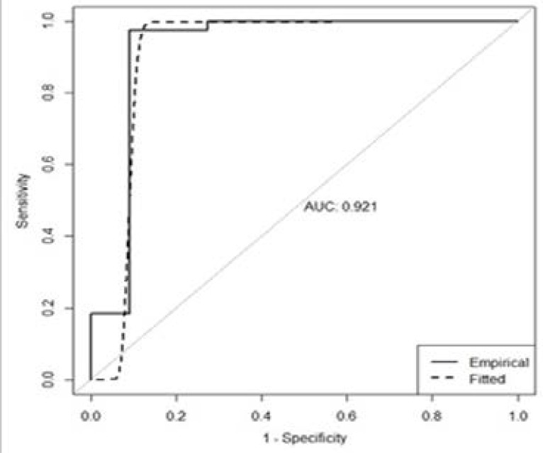
Results: Of the 72 liver masses, 51 were benign and 21 were malignant. Benign lesions comprised 38 hemangiomas and 13 simple cysts. Malignant lesions comprised 9 hepatocellular carcinomas, and 12 metastases. The highest ADC values were measured for cysts (3.782±0.53×10(-3) mm(2)/s) and hemangiomas (2.705±0.63×10(-3) mm(2)/s). The average ADC value of hemangiomas was significantly higher than malignant lesions and the normal control group (p<0.001). The average ADC value of cysts were significantly higher when compared to hemangiomas and normal control group (p<0.001). To distinguish hemangiomas from malignant liver lesions, the "cut-off" ADC value of 1.800×10(-3) mm(2)/s had a sensitivity of 97.4% and a specificity of 90.9%. To distinguish hemangioma from normal liver parenchyma the "cut-off" value of 1.858×10(-3) mm(2)/s had a sensitivity of 97.4% and a specificity of 95.7%. To distinguish benign liver lesions from malignant liver lesions the "cut-off" value of 1.800×10(-3) mm(2)/s had a sensitivity of 96.1% and a specificity of 90.0%.
Conclusion: DWI and quantitative measurement of ADC values can be used in differential diagnosis of benign and malignant liver lesions and also in the diagnosis and differentiation of hemangiomas. When dynamic examination cannot distinguish cases with vascular metastasis and lesions from hemangioma, DWI and ADC values can be useful in the primary diagnosis and differential diagnosis. The technique does not require contrast material, so it can safely be used in patients with renal failure.
Keywords: Liver; apparent diffusion coefficient; diffusion weighted magnetic resonance imaging; hemangioma.
Figures
Similar articles
-
Differentiation between cavernous hemangiomas and untreated malignant neoplasms of the liver with free-breathing diffusion-weighted MR imaging: comparison with T2-weighted fast spin-echo MR imaging.Eur J Radiol. 2011 Nov;80(2):316-24. doi: 10.1016/j.ejrad.2010.08.011. Epub 2010 Aug 30. Eur J Radiol. 2011. PMID: 20800983
-
The value of diffusion-weighted imaging in characterizing focal liver masses.Acad Radiol. 2009 Oct;16(10):1208-14. doi: 10.1016/j.acra.2009.05.013. Epub 2009 Jul 15. Acad Radiol. 2009. PMID: 19608435
-
Characterizing focal hepatic lesions by free-breathing intravoxel incoherent motion MRI at 3.0 T.Acta Radiol. 2014 Dec;55(10):1166-73. doi: 10.1177/0284185113514966. Epub 2013 Dec 6. Acta Radiol. 2014. PMID: 24316660
-
Lesion discrimination with breath-hold hepatic diffusion-weighted imaging: a meta-analysis.World J Gastroenterol. 2015 Feb 7;21(5):1621-7. doi: 10.3748/wjg.v21.i5.1621. World J Gastroenterol. 2015. PMID: 25663782 Free PMC article. Review.
-
Diagnostic accuracy of apparent diffusion coefficient (ADC) value in differentiating malignant from benign solid liver lesions: a systematic review and meta-analysis.Br J Radiol. 2021 Jul 1;94(1123):20210059. doi: 10.1259/bjr.20210059. Epub 2021 Jun 16. Br J Radiol. 2021. PMID: 34111960 Free PMC article.
Cited by
-
Evaluation of ADCratio on liver MRI diffusion to discriminate benign versus malignant solid liver lesions.Eur J Radiol Open. 2018 Nov 15;5:209-214. doi: 10.1016/j.ejro.2018.10.002. eCollection 2018. Eur J Radiol Open. 2018. PMID: 30480057 Free PMC article.
-
An explanation for the triphasic dependency of apparent diffusion coefficient (ADC) on T2 relaxation time: the multiple T2 compartments model.Quant Imaging Med Surg. 2025 Apr 1;15(4):3779-3791. doi: 10.21037/qims-2025-195. Epub 2025 Mar 5. Quant Imaging Med Surg. 2025. PMID: 40235750 Free PMC article. No abstract available.
-
Evaluation of the Effect of Patient Preparation Using Castor Oil on ADC Value of Focal Liver Lesion.Int J Gen Med. 2021 Feb 16;14:469-474. doi: 10.2147/IJGM.S289661. eCollection 2021. Int J Gen Med. 2021. PMID: 33623419 Free PMC article.
-
Diffusion Kurtosis MR Imaging versus Conventional Diffusion-Weighted Imaging for Distinguishing Hepatocellular Carcinoma from Benign Hepatic Nodules.Contrast Media Mol Imaging. 2019 Jul 17;2019:2030147. doi: 10.1155/2019/2030147. eCollection 2019. Contrast Media Mol Imaging. 2019. PMID: 31396023 Free PMC article.
-
Updates on communicable and non-communicable diseases in LMICs.Afr Health Sci. 2016 Mar;16(1):i-iv. doi: 10.4314/ahs.v16i1.. Afr Health Sci. 2016. PMID: 27358652 Free PMC article. No abstract available.
References
-
- Ichikawa T, Haradome H, Hachiya J, Nitatori T, Araki T. Diffusion-weighted MR imaging with single-shot echo-planar imaging in the upper abdomen: preliminary clinical experience in 61 patients. Abdom Imaging. 1999;24:456–461. - PubMed
-
- Moteki T, Horikoshi H, Oya N, Aoki J, Endo K. Evaluation of hepatic lesions and hepatic parenchyma using diffusion-weighted reordered turboFLASH magnetic resonance images. J Magn Reson Imaging. 2002;15:564–572. - PubMed
-
- Warach S, Chien D, Li W, Ronthal M, Edelman RR. Fast magnetic resonance diffusion-weighted imaging of acute human stroke. Neurology. 1992;42:1717–1723. - PubMed
-
- Back T, Hoehn-Berlage M, Kohno K, Hossmann KA. Diffusion nuclear magnetic resonance imaging in experimental stroke: correlation with cerebral metabolites. Stroke. 1994;25:494–500. - PubMed
-
- Lutsep HL, Albers GW, DeCrespigny A, Kamat GN, Marks MP, Moseley ME. Clinical utility of diffusion-weighted magnetic resonance imaging in the assessment of ischemic stroke. Ann Neurol. 1997;41:574–580. - PubMed
MeSH terms
LinkOut - more resources
Full Text Sources
Other Literature Sources
Medical