

Solitary fibrous tumor of the pancreas: Case report and review of the literature
- PMID: 27358679
- PMCID: PMC4919714
- DOI: 10.4240/wjgs.v8.i6.461
Solitary fibrous tumor of the pancreas: Case report and review of the literature
Abstract
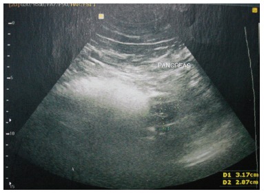
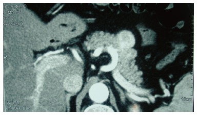
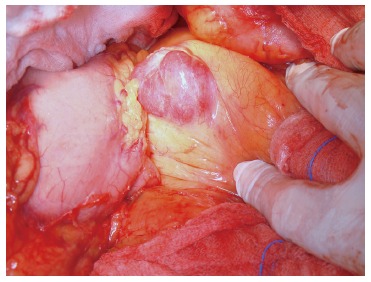
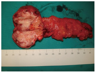
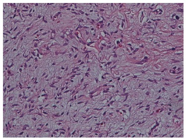
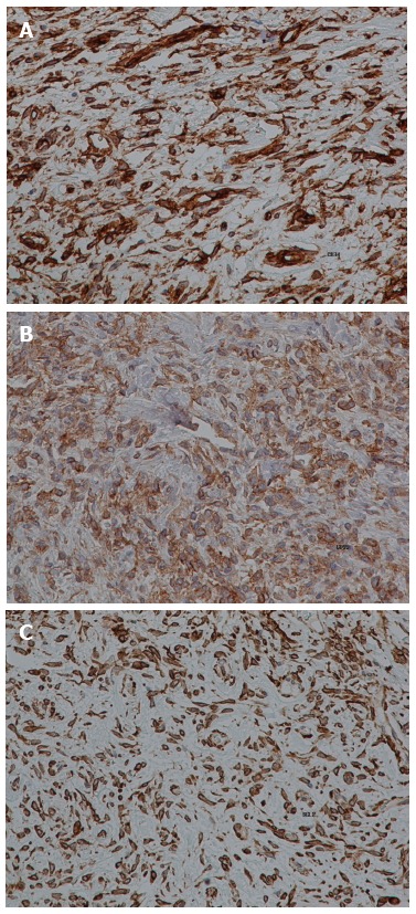
Solitary fibrous tumor (SFT) is a mesenchymal tumor typically located in the pleura, but can also be found as an asymptomatic mass in other areas, including the liver, peritoneum, kidney and salivary glands. However, SFT rarely locates in the pancreas. We present such a case of pancreatic SFT, along with a review of all reported cases. A 55-year-old man was treated surgically for an asymptomatic pancreatic mass after a rigorous preoperative control. Histologic examination of the resected specimen showed characteristics of an SFT. As only 15 cases of pancreatic SFT have been reported so far, an attempt to compare the cases was considered intriguing. We found that patients with pancreatic SFT were mainly women (81.25%), with a median age of 54 years at the time of diagnosis and a median tumor size of 5.83 cm. Pancreatic SFTs were revealed incidentally in 50% of cases, and all of them showed an enhancement through arterial computed tomography. All tumors were positive for CD34, ten were positive for Bcl-2, and twelve were negative for S100. The diagnosis of this pancreatic tumor is established by a combination of clinical suspicion, imaging procedures and histological findings, and is confirmed by immunohistochemical staining. Although the behavior of SFTs is rather benign, close clinical follow-up is recommended due to a potentially malignant nature.
Keywords: Differential diagnosis; Mesenchymal tumors; Pancreas; Solitary fibrous tumor; Solitary fibrous tumor treatment.
Figures
References
-
- Tasdemir A, Soyuer I, Yurci A, Karahanli I, Akyildiz H. A huge solitary fibrous tumor localized in the pancreas: a young women. JOP. 2012;13:304–307. - PubMed
-
- Ginat DT, Bokhari A, Bhatt S, Dogra V. Imaging features of solitary fibrous tumors. AJR Am J Roentgenol. 2011;196:487–495. - PubMed
-
- Chetty R, Jain R, Serra S. Solitary fibrous tumor of the pancreas. Ann Diagn Pathol. 2009;13:339–343. - PubMed
-
- Lüttges J, Mentzel T, Hübner G, Klöppel G. Solitary fibrous tumour of the pancreas: a new member of the small group of mesenchymal pancreatic tumours. Virchows Arch. 1999;435:37–42. - PubMed
-
- Srinivasan VD, Wayne JD, Rao MS, Zynger DL. Solitary fibrous tumor of the pancreas: case report with cytologic and surgical pathology correlation and review of the literature. JOP. 2008;9:526–530. - PubMed
Publication types
LinkOut - more resources
Full Text Sources
Other Literature Sources
