. 2017 Feb;219(2):521-536.
doi: 10.1111/apha.12742. Epub 2016 Jul 20.

Claudin-2-mediated cation and water transport share a common pore

Affiliations

Claudin-2-mediated cation and water transport share a common pore

R Rosenthal et al. Acta Physiol (Oxf). 2017 Feb.

Abstract

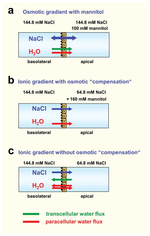
Aim: Claudin-2 is a tight junction protein typically located in 'leaky' epithelia exhibiting large paracellular permeabilities like small intestine and proximal kidney tubule. Former studies revealed that claudin-2 forms paracellular channels for small cations like sodium and potassium and also paracellular channels for water. This study analyses whether the diffusive transport of sodium and water occurs through a common pore of the claudin-2 channel.

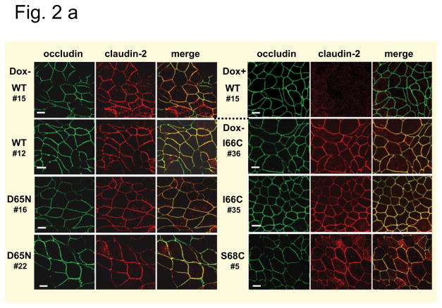
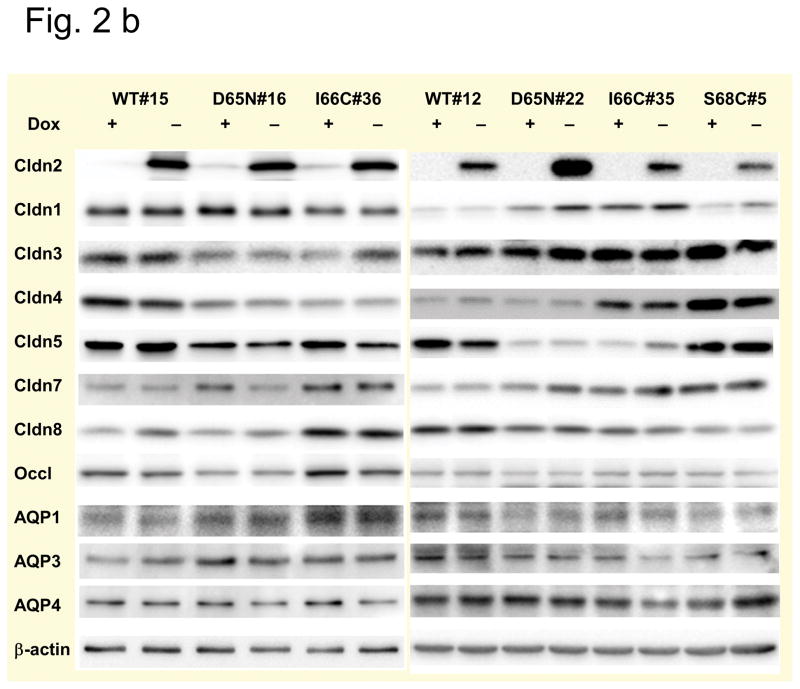
Methods: Wild-type claudin-2 and different claudin-2 mutants were expressed in MDCK I kidney tubule cells using an inducible system. Ion and water permeability and the effect of blocking reagents on both were investigated on different clones of the mutants.

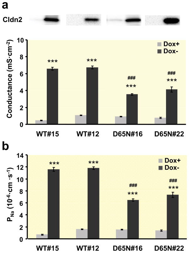
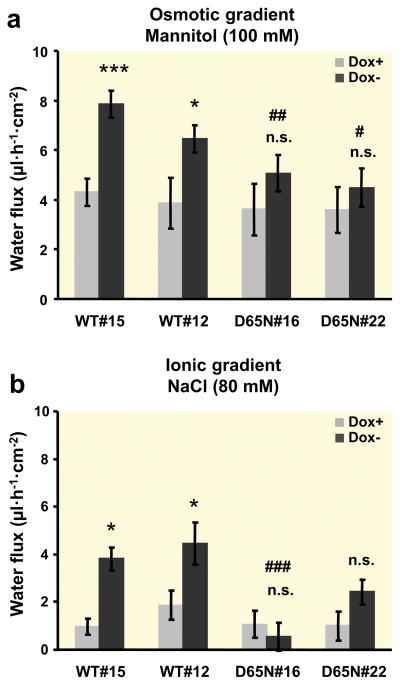
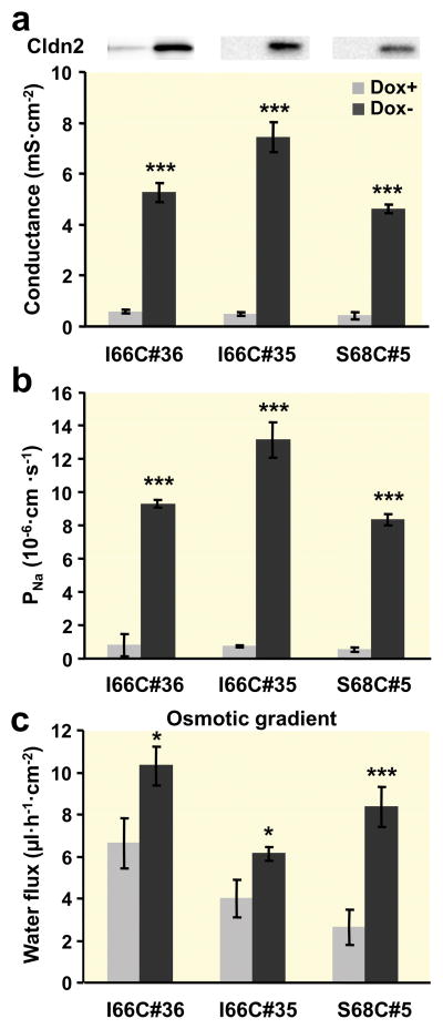
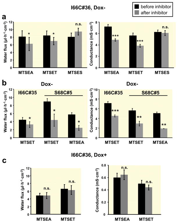
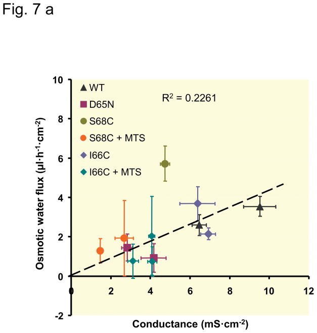
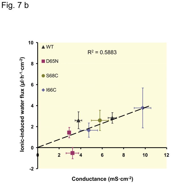
Results: Neutralization of a negatively charged cation interaction site in the pore with the mutation, D65N, decreased both sodium permeability and water permeability. Claudin-2 mutants (I66C and S68C) with substitution of the pore-lining amino acids with cysteine were used to test the effect of steric blocking of the claudin-2 pore by thiol-reactive reagents. Addition of thiol-reactive reagents to these mutants simultaneously decreased conductance and water permeability. Remarkably, all experimental perturbations caused parallel changes in ion conductance and water permeability, disproving different or independent passage pathways.

Conclusion: Our results indicate that claudin-2-mediated cation and water transport are frictionally coupled and share a common pore. This pore is lined and determined in permeability by amino acid residues of the first extracellular loop of claudin-2.

Keywords: claudin-2 pore; common pore for sodium and water; paracellular transport.


Conflict of interest statement

Declaration The authors declare that they have no conflict of interest. The authors confirm that the material submitted is conform with Good Publishing Practice in Physiology 2013: Guidelines for Acta Physiologica (Persson PB, 2013).

Figures

Fig. 1
Fig. 1. Experimental conditions for measuring water flow
For measuring water flux the Ussing chamber was filled with HEPES buffered solution with a total NaCl concentration of 144.8 mM. The solution in the apical compartment was changed according to the following conditions: (a) Osmotic gradient with mannitol in the apical compartment. The osmotic gradient with mannitol in the basolateral compartment is not shown here. (b) Ionic gradient with the high NaCl concentration in the basolateral compartment and osmotic “compensation” by mannitol in the apical compartment. As MDCK cells are unable to secrete Na+, no transcellular Na+ flux will occur under these conditions. (c) Ionic gradient with the high NaCl concentration in the basolateral compartment and no osmotic “compensation”. As in (b) no transcellular Na+ flux will occur under these conditions. The osmotic and ionic gradients aredirected conversely. Assuming that claudin-2 is the only paracellular water channel, paracellular water flux induced by the ionic gradient will only occur in claudin-2-expressing MDCK I cells. During all experiments the transepithelial potential was clamped at 0 mV. This might favour an isolated paracellular Na+ passage in claudin-2-expressing cells and thus induce paracellular water flux.
Fig. 2
Fig. 2. Tight junction proteins and AQPs in MDCK I TetOff cells stably transfected with mouse wild-type and mutant claudin-2
(a) Subcellular localization of claudin-2 in the cells. Different clones expressing wild-type (WT) claudin-2, and the claudin-2 mutants D65N, and the cysteine mutants I66C and S68C were induced to express claudin-2 (Dox−) and immunofluorescence stained using an antibody against claudin-2 (left) and the tight junction marker occludin (middle). The colocalization of both proteins was confirmed by the merged view (right). No claudin-2 signal could be detected in the uninduced (Dox+) cells, here shown for the uninduced cells of wild-type claudin-2, clone 15. Bar 10 μm. (b) Claudin and AQP expression in the cells. Western blot analysis showing the expression of the transfected claudin-2 proteins, endogenous claudins, and aquaporins (AQP) in uninduced (Dox+) and induced (Dox−) MDCK I TetOff cells transfected with wild-type (WT) claudin-2 and the claudin-2 mutants.
Fig. 3
Fig. 3. Electrophysiological properties of MDCK I TetOff cells expressing wild-type or D65N claudin-2
(a) Conductance and (b) Na+ permeability of two different clones of wild-type (WT) and mutated (D65N) claudin-2 (n=7–14). The upper panel in (A) is a Western blot (from Fig. 2b) showing claudin-2 protein expression in each clone (left lane, Dox+; right lane, Dox−). Permeability was derived from measurement of dilution potentials and calculated by means of the Goldman-Hodgkin-Katz equation. As expected, a strong difference in conductance and permeability could be observed between induced (Dox−) and uninduced (Dox+) cells and also between cells expressing wild-type and mutated claudin-2. (*** p < 0.001 vs. Dox+, ### p < 0.001 vs. WT#12 and WT#15).
Fig. 4
Fig. 4. Water flux across layers of MDCK I TetOff cells expressing wild-type or D65N claudin-2
(a) Water flux induced by an osmotic gradient with 100 mM mannitol on the apical side of the cell layer (n=7–12). (b) Water flux induced by an ionic gradient with a difference of 80 mM NaCl and the high NaCl concentration at the basolateral side of the cell layer. The osmotic difference was compensated by the addition of 160 mM mannitol to the apical side (n=6–13). Positive water flux is defined as flow from basolateral to apical side. In the wild-type claudin-2-transfected cells, water flux was increased in the induced Dox− cells compared to the uninduced Dox+ cells under both osmotic and ionic gradients, whereas no difference could be observed in D65N claudin-2-transfected cells. (*p < 0.05, ***p < 0.001, n.s. = not significant vs. Dox+; # p < 0.05 vs. WT#12 and WT#15, ## p < 0.01 vs. WT#15, ### p < 0.001 vs. WT#15 and WT#12).
Fig. 5
Fig. 5. Characterization of MDCK I TetOff cells stably transfected with claudin-2 cysteine mutants
(a) Conductance, (b) Na+ permeability, and (c) osmotic gradient-induced water flux (100 mM mannitol at the apical side) of two different clones of I66C and one clone of S68C claudin-2 mutants (n=7–14). The upper panel in (A) is a Western blot (from Fig. 2b) showing claudin-2 protein expression in each clone (left lane, Dox+; right lane, Dox−). Permeability was derived from measurement of dilution potentials and calculated by means of the Goldman-Hodgkin-Katz equation. Conductance, Na+ permeability, and water flux were all increased in induced (Dox−) cells. (* p < 0.05, *** p < 0.001 vs. Dox+).
Fig. 6
Fig. 6. Effects of MTS reagents on water flux and conductance in MDCK I TetOff cells expressing claudin-2 cysteine mutants
Osmotic gradient-induced water transport (left panel) and conductance (right panel) before and after addition of different MTS reagents (“inhibitor”) in induced (Dox−, a, b) and uninduced (Dox+, c) MDCK I TetOff cells expressing I66C claudin-2 (#36), and in induced (Dox−, b) cells expressing I66C (#35) and S68C (#5) claudin-2 (n=4–10). MTSEA (2.5 mM) and MTSET (1 mM) caused a decrease in water flux and conductance in Dox− cells of all mutants, whereas MTSES (5 mM) had no effect on either conductance or water flux. MTSEA and MTSET did not exert any effect on Dox+ cells. (n.s. = not significant, * p < 0.05, ** p < 0.01, *** p < 0.001 vs. before inhibitor, paired Student’s t test).
Fig. 7
Fig. 7. Relationship between claudin-2-mediated water flux and claudin-2-mediated conductance
The water flux mediated by claudin-2 was calculated as the difference between water flux of uninduced (Dox+) and induced (Dox−) cells. Claudin-2 conductance was calculated as the difference between conductance of uninduced (Dox+) and induced (Dox−) cells. Each data point represents the values (mean ± SEM) determined in one clonal cell line. Decrease in conductance due to charge neutralization (D65N), blocking of the claudin-2 pore with MTS reagents (only in a), or clonal variation, are all associated with a commensurate decrease in water flux. The dashed line was fitted by linear regression to all data points. (a) Relationship between osmotic-induced water flux and conductance, slope 0.336 μl·h−1·mS−1. (b) Relationship between water flux induced by the NaCl gradient (ionic-induced water flux) and conductance, slope 0.436 μl·h−1·mS−1.

References

    1. Agre P, Preston GM, Smith BL, Jung JS, Raina S, Moon C, Guggino WB, Nielsen S. Aquaporin CHIP: the archetypal molecular water channel. Am J Physiol. 1993;265:463–76. - PubMed
    1. Amasheh S, Meiri N, Gitter AH, Schöneberg T, Mankertz J, Schulzke JD, Fromm M. Claudin-2 expression induces cation-selective channels in tight junctions of epithelial cells. J Cell Sci. 2002;115:4969–76. - PubMed
    1. Angelow S, El-Husseini R, Kanzawa SA, Yu AS. Renal localization and function of the tight junction protein, claudin-19. Am J Physiol Renal Physiol. 2007;293:166–77. - PubMed
    1. Angelow S, Yu AS. Structure-function studies of claudin extracellular domains by cysteine-scanning mutagenesis. J Biol Chem. 2009;284:29205–17. - PMC - PubMed
    1. Bello-Reuss E. Cell membranes and paracellular resistances in isolated renal proximal tubules from rabbit and Ambystoma. J Physiol. 1986;370:25–38. - PMC - PubMed

Publication types