. 2016 Jul 7;535(7610):182-6.
doi: 10.1038/nature18324. Epub 2016 Jun 29.

Allosteric coupling from G protein to the agonist-binding pocket in GPCRs

Allosteric coupling from G protein to the agonist-binding pocket in GPCRs

Brian T DeVree et al. Nature. .

Abstract

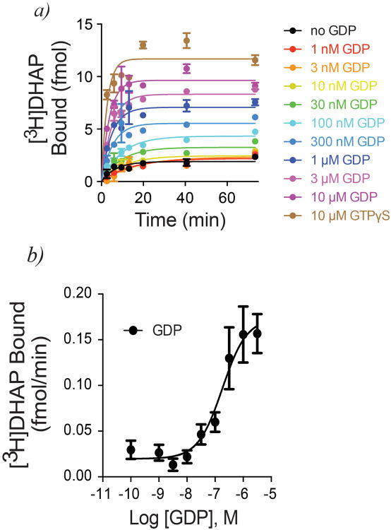
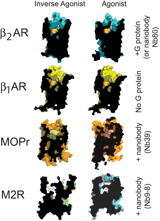
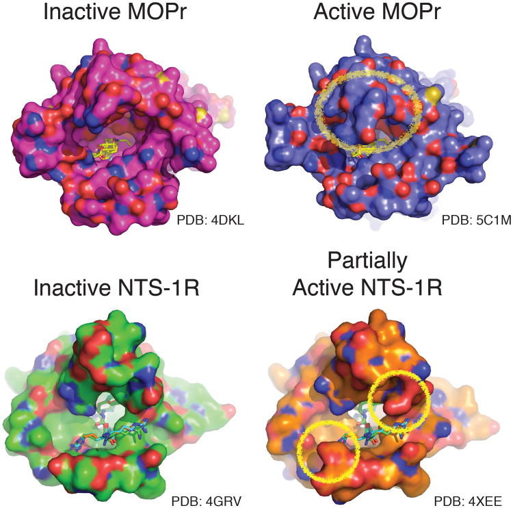
G-protein-coupled receptors (GPCRs) remain the primary conduit by which cells detect environmental stimuli and communicate with each other. Upon activation by extracellular agonists, these seven-transmembrane-domain-containing receptors interact with heterotrimeric G proteins to regulate downstream second messenger and/or protein kinase cascades. Crystallographic evidence from a prototypic GPCR, the β2-adrenergic receptor (β2AR), in complex with its cognate G protein, Gs, has provided a model for how agonist binding promotes conformational changes that propagate through the GPCR and into the nucleotide-binding pocket of the G protein α-subunit to catalyse GDP release, the key step required for GTP binding and activation of G proteins. The structure also offers hints about how G-protein binding may, in turn, allosterically influence ligand binding. Here we provide functional evidence that G-protein coupling to the β2AR stabilizes a ‘closed’ receptor conformation characterized by restricted access to and egress from the hormone-binding site. Surprisingly, the effects of G protein on the hormone-binding site can be observed in the absence of a bound agonist, where G-protein coupling driven by basal receptor activity impedes the association of agonists, partial agonists, antagonists and inverse agonists. The ability of bound ligands to dissociate from the receptor is also hindered, providing a structural explanation for the G-protein-mediated enhancement of agonist affinity, which has been observed for many GPCR–G-protein pairs. Our data also indicate that, in contrast to agonist binding alone, coupling of a G protein in the absence of an agonist stabilizes large structural changes in a GPCR. The effects of nucleotide-free G protein on ligand-binding kinetics are shared by other members of the superfamily of GPCRs, suggesting that a common mechanism may underlie G-protein-mediated enhancement of agonist affinity.


Figures

Extended Data Figure 1
Extended Data Figure 1. Confirmation of nucleotide removal from β2AR•Gs by apyrase
Gs and Flag-tagged β2AR were reconstituted in rHDL and treated with the non-specific nucleotide lyase, apyrase. Samples were applied to an anti-Flag affinity resin to remove products of the GDP degradation (GMP and Pi). Samples were incubated with 100 nM [35S]GTPγS at room temperature. At various times, samples were subjected to rapid filtration through glass fiber filters (GF/B) followed by 10 volumes of ice-cold buffer washes containing 10 μM GDP. Filters were dried and subjected to liquid scintillation counting (Top-Count™, Perkin-Elmer). To a first approximation the rapid binding event suggests that the complex is empty of nucleotide, based on the limited temporal resolution of this mixing and filtration technique. [3H]DHAP and [35S]GTPγS binding to the reconstituted complex yields a final R:G ratio of 1:0.95, suggesting that up to 95% of the β2AR-rHDL particles contain a single functional G protein. This suggest that only those G proteins associated with the β2AR will bind [35S]GTPγS within this time frame in the absence of receptor agonists. Data are shown as mean ± SEM from n=3 independent experiments performed in duplicate.
Extended Data Figure 2
Extended Data Figure 2. GDP accelerates [3H]DHAP binding to β2AR•Gs
a) Time course monitoring [3H]DHAP association to apyrase-treated β2AR•Gs complexes in the presence of varying GDP concentrations. GDP increases both the observed association rate constant and the maximum binding of [3H]DHAP. b) Concentration-response showing enhancement of the observed [3H]DHAP association rate constant by GDP (EC50 = 181 ± 66 nM). All data are shown as mean ± SEM from n=3 independent experiments performed in duplicate.
Extended Data Figure 3
Extended Data Figure 3. Effect of guanine nucleotides on [3H]DHAP binding to β2AR•Gs
a) In saturation binding assays, addition of GTPγS to apyrase-treated β2AR•Gs complexes increased the observed Bmax for [3H]DHAP without significantly altering Kd (Control: Bmax = 5.5 ± 0.52 fmol, Kd = 0.88 nM; +GTPγS: Bmax = 16.6 ± 1.9 fmol, Kd = 0.56 nM) b) Both GDP and GTPγS could enhance maximal [3H]DHAP binding in a concentration-dependent manner (GDP Log(EC50) = -6.42 ± 0.12, or EC50 ∼386 nM; GTPγS Log(EC50) = -7.45 ± -0.16, or EC50 ∼35 nM). All data are shown as mean ± SEM from n=3 independent experiments performed in duplicate.
Extended Data Figure 4
Extended Data Figure 4. Effect of Nb80 on antagonist binding to β2AR
a) Association of [3H]DHAP is progressively slowed following pre-incubation of β2AR with increasing concentrations of Nb80. b) If [3H]DHAP is allowed to first equilibrate with β2AR, Nb80 slows [3H]DHAP dissociation from β2AR in a concentration-dependent manner. c) Due to the dramatic slowing of [3H]DHAP binding kinetics, Nb80 (but not a control nanobody, Nb30, which has no effect on agonist affinity for β2AR) appears competitive with [3H]DHAP if insufficient time is given to reach equilibrium. Data shown are from assays incubated 90 minutes at room temperature. All data are shown as mean ± SEM from n=3 independent experiments performed in duplicate.
Extended Data Figure 5
Extended Data Figure 5. Y308A mutation abolishes the rate-slowing effects of Nb80
a) and b) Time course of [3H]DHAP binding to wild-type β2AR (a) or β2AR-Y308A (b) following pre-incubation of receptor with Nb80. Nb80 significantly slowed [3H]DHAP association to wild-type β2AR (-Nb80 kobs = 0.45 ± 0.05 min-1 or t½ = 1.5 ± 0.2 min, +Nb80 kobs = 0.20 ± 0.03 min-1 or t½ = 3.5 ± 0.5 min; p = 0.011 by an unpaired two-tailed t-test), but less effectively slowed [3H]DHAP association to β2AR-Y308A (-Nb80 kobs = 0.50 ± 0.06 min-1 or t½ = 1.4 ± 0.2 min; +Nb80 kobs = 0.32 ± 0.01 min-1 or t½ = 2.2 ± 0.1 min; p = 0.05 by an unpaired two-tailed t-test. All data are shown as mean ± SEM from n=4 (-Nb80) or n=3 (+Nb80) independent experiments performed in duplicate. c) and d) Time course of [3H]formoterol binding to wild-type β2AR (c) or β2AR-Y308A (d) following pre-incubation of receptor with Nb80. Nb80 slowed [3H]formoterol association to wild-type β2AR (0.1 μM Nb80 kobs = 0.68 ± 0.13 min-1 or t½ = 1.0 ± 0.2 min, 10 μM Nb80 kobs = 0.27 ± 0.05 min-1 or t½ = 2.6 ± 0.5 min; p = 0.031 by an unpaired two-tailed t-test). However, with β2AR-Y308A, Nb80 had little effect on the observed association rate constant but enhanced the amount of [3H]formoterol bound (0.1 μM Nb80 kobs = 0.37 ± 0.11 min-1 or t½ = 1.9 ± 0.6 min with a plateau of 10.1 ± 0.8 fmol, 10 μM Nb80 kobs = 0.53 ± 0.13 min-1 or t½ = 1.3 ± 0.4 min with a plateau of 21.3 ± 1.2 fmol; unpaired two-tailed t-test of the kobs values showed p = 0.4). All data are shown as mean ± SEM from n=4 independent experiments performed in duplicate.
Extended Data Figure 6
Extended Data Figure 6. The closed conformation stabilized by agonist and G protein (or mimic)
Illustrated are the crystal structures of agonist- vs. inverse agonist-bound of the β2AR (cyan) and β1AR (yellow), where only β2AR is bound to G protein. Similarly, the μ-opioid receptor (MOPr, orange) adopts a closed conformation upon binding G protein surrogate, Nb39. (β2AR; PDB 2RH1, β2AR•Gs; PDB 3SN6, β1AR; PDB 2YCW, β1AR-iso; PDB 2Y03, MOPr; PDB 4DKL, MOPr-Nb39; PDB 5C1M, M2R; PDB 3UON, M2R-Nb9-8; PDB 4MQS).
Extended Data Figure 7
Extended Data Figure 7. Effect of guanine nucleotides on [3H]antagonist binding are also seen in competition binding assays
a) Agonist (isoproterenol) competition binding using apyrase-treated β2AR•Gs complexes shows the characteristic G protein-dependent shift in agonist affinity, along with a dramatic increase in total [3H]DHAP binding, upon the addition of 10 μM GTPγS. b) Normalization of the data from a) yields a plot representative of what is commonly reported in the literature. c) Similar to β2AR, agonist (morphine) competition binding using MOPr•Go complexes shows the characteristic G protein-dependent shift in agonist affinity, along with a dramatic increase in total [3H]DPN binding, upon the addition of 10 μM GTPγS. d) Normalization of the data from c). All data are shown as mean ± SEM from n=3 independent experiments performed in duplicate.
Extended Data Figure 8
Extended Data Figure 8. Mu opioid receptor and M2 muscarinic acetylcholine receptor behave similarly to β2AR when bound to nucleotide-free G protein or an active-state stabilizing nanobody
a) Following apyrase treatment of M2R•Go complexes, addition of 10 μM GTPγS enhances association of [3H]N-methylscopolamine ([3H]NMS) to M2R (Vehicle kobs = 0.32 ± 0.02 min-1 or t½ = 2.2 ± 0.1 min, +GTPγS kobs = 0.54 ± 0.02 min-1 or t½ = 1.3 ± 0.1 min; p = 0.002 by an unpaired two-tailed t-test). Data are shown as mean ± SEM from n=3 independent experiments performed in duplicate. Addition of GDP was also able to increase the rate of [3H]NMS binding (inset; pEC50 = 6.91 ± 0.18 or EC50 ∼123 nM; mean ± SEM from n=2 independent experiments performed in duplicate). b) Pre-treatment of M2R with either 10 μM (black circles) or 100 μM (red squares) Nb9-827 impairs association of [3H]iperoxo to M2R (10 μM Nb9-8 kobs = 0.68 ± 0.09 min-1 or t½ = 1.0 ± 0.2 min, 100 μM Nb9-8 kobs = 0.25 ± 0.04 min-1 or t½ = 2.8 ± 0.5 min; p = 0.04 by an unpaired two-tailed t-test). Data are shown as mean ± SEM from n=3 (10 μM Nb9-8) or n=2 (100 μM Nb9-8) independent experiments performed in duplicate. c) Addition of 10 μM GTPγS to apyrase-treated MOPr•Go complexes hastened association of the antagonist [3H]diprenorphine ([3H]DPN) to MOPr (Apyrase kobs = 0.06 ± 0.02 min-1 or t½ = 9.8 ± 1.3 min, +GTPγS kobs = 0.12 ± 0.01 min-1 or t½ = 5.6 ± 0.6 min; p = 0.1 by an unpaired two-tailed t-test). The effect of nucleotide-free G protein was recapitulated by pre-incubating MOPr with Nb39 (inset; control kobs = 0.13± 0.01 min-1, +100 μM Nb39 kobs = 0.07 ± 0.02 min-1). Data are shown as mean ± SEM from n=2 (MOPr•Go) or n=3 (MOPr + Nb39) independent experiments performed in duplicate.
Extended Data Figure 9
Extended Data Figure 9. The extracellular regions in the active conformations of peptide hormone/agonist receptors MOPr and NTS-R1
Illustrated are the crystal structures of the inactive and active (or partially active NTS-R1) conformations of the MOPr and NTS-R1 from the top or extracellular view of the receptor. The surface rendering highlights residues or structure on the extracellular face that change upon receptor activation (circled). The mu-opioid receptor (MOPr) in its inactive conformation (purple) is compared to the Nb39-bound (G protein mimic) form in blue. Similarly, the inactive NTS-R1 (green) is compared with a mutant NTS-R1 that adopts a partially active conformation (orange). (MOPr; PDB 4DKL, MOPr-Nb39; PDB 5C1M, NTS-R1; PDB 4GRV and active-like NTS-R1; PDB 4XEE).
Extended Data Figure 10
Extended Data Figure 10. Model of G protein-dependent high-affinity agonist binding
As in Figure 5 (of the main text), nucleotide-free G protein-stabilized family A GPCRs experience alterations in the extracellular face of the receptor, thus affecting orthosteric binding site. In a monoamine receptor like the β2AR, G protein binding and GDP loss accompanies the stabilization of a closed, active conformation of the receptor, as in a). b) For family members such as MOPr or NTS-R1, where the peptide hormones/agonists are considerably larger, the influence of the G protein-mediated changes in the extracellular domain structure result in similar effects on orthosteric ligand dissociation. Rather than closing over the orthosteric site as with monoamine receptors as in a) the extracellular face may contain structures and residues that ‘pinch’ the larger ligands.
Figure 1
Figure 1. Guanine nucleotides influence antagonist binding to β2AR•Gs complexes
a) Binding of 2 nM [3H]DHAP to β2AR•Gs in the absence or presence of GDP. Addition of apyrase to GDP-bound β2AR•Gs led to a progressive decrease in [3H]DHAP binding over time, which could be restored with excess GDP. b) Addition of increasing concentrations of GDP enhances the rate and extent of [3H]DHAP binding to apyrase-treated β2AR•Gs complexes. Data in a) are shown as mean ± SEM from n=3 independent experiments performed in duplicate. Data in b) are representative of three independent experiments.
Figure 2
Figure 2. Trapping active-state β2AR with Nb80 slows both antagonist and agonist association
a) Nb80 (red) mimics G protein (yellow) in both its binding site and the β2AR conformation it stabilizes. The structure of Nb80-bound β2AR (3p0g) is shown in orange, Gs-bound β2AR (3sn6) in cyan. b) Pre-incubation of β2AR with increasing concentrations of Nb80 progressively slows association of neutral antagonist [3H]DHAP to β2AR. c) Nb80 also slows association of full agonist [3H]formoterol, d) partial agonist [3H]CGP12177, and e) inverse agonist [3H]carvedilol to β2AR. f) Nb80 stabilizes the closed, active conformation and slows [3H]DHAP dissociation from β2AR in a concentration-dependent manner. Data in b) and f) are representative of three independent experiments. All other data are specific binding, shown as mean ± SEM from n=3 independent experiments performed in duplicate.
Figure 3
Figure 3. Activation of β2AR closes the hormone binding site
a) Stabilization of the β2AR active conformation by Gs (or Nb80) brings the side chains of Phe193ECL2 and Tyr3087.35 closer to one another compared to their positions in structures in the absence of G protein. b) Closer view of the orthosteric site, highlighting Phe193ECL2 and Tyr3087.35. Distances (in Ångstroms) between the hydroxyl on Tyr3087.35 and 2-carbon on the phenyl ring of Phe193ECL2 are indicated. c) and d) A surface view comparing the extracellular face of β2AR in inactive (panel c) or active (panel d) conformations, showing how G protein-stabilized structural rearrangements occlude the hormone binding site in the active state. e) and f) Cutaway view illustrating closure of the hormone binding site around the bound agonist in the active state. The inverse agonist carazolol is shown in orange, the agonist BI-167107 is shown in yellow.
Figure 4
Figure 4. Allosteric communication between β2AR G protein- and hormone-binding sites
a) In the β2AR active state (cyan), the cytoplasmic end of TM6 moves away from the receptor core by ∼14 Å relative to its position in the inactive-state structure, allowing for an inward movement of TM7. b) Rotation of TM7 allows Tyr3267.53 (of the highly conserved NPxxY motif) to fill the space vacated by the conserved aliphatic residue Ile2786.40. c) The rotation of TM7 repositions Tyr3087.35 and Lys3057.32. This conformational change allows Lys3057.32 to coordinate the backbone carbonyl of Phe193ECL2, stabilizing its movement toward Tyr3087.35 to form a “lid” over the hormone binding site.
Figure 5
Figure 5. Basis for G protein-dependent high-affinity agonist binding
Agonist binding promotes the receptor-G protein interaction and GDP release from Gα. In this nucleotide-free state, the C-terminal helix of Gα remains embedded in the receptor core, stabilizing the conformational changes at both the intracellular and extracellular faces of the receptor. At the extracellular side, the orthosteric binding site closes around the bound agonist, sterically opposing agonist dissociation and thereby enhancing the observed affinity. Constitutive (basal) receptor activity may also activate the G protein, releasing GDP and thereby stabilizing the ‘closed’ conformation of the receptor in the absence of an agonist. See also Extended Data Figure 10.

Similar articles

Cited by

References

    1. Pierce KL, 1, Premont RT, Lefkowitz RJ. Seven-transmembrane receptors. Nat Rev Mol Cell Biol. 2002 Sep;3(9):639–50. - PubMed
    1. Rasmussen SG, et al. Crystal structure of the β2-adrenergic receptor–Gs protein complex. Nature. 2011;477:549–555. - PMC - PubMed
    1. Venter JC, et al. The sequence of the human genome. Science. 2001;291:1304–51. - PubMed
    1. Sprang SR. G protein mechanisms: insights from structural analysis. Annu Rev Biochem. 1997;66:639–78. - PubMed
    1. Chung K, et al. Conformational changes in the G protein Gs induced by the β2-adrenergic receptor. Nature. 2011;477:611–615. - PMC - PubMed

Publication types

MeSH terms