. 2016 Jul 7;535(7610):173-7.
doi: 10.1038/nature18317. Epub 2016 Jun 29.

A core viral protein binds host nucleosomes to sequester immune danger signals

A core viral protein binds host nucleosomes to sequester immune danger signals

Daphne C Avgousti et al. Nature. .

Abstract

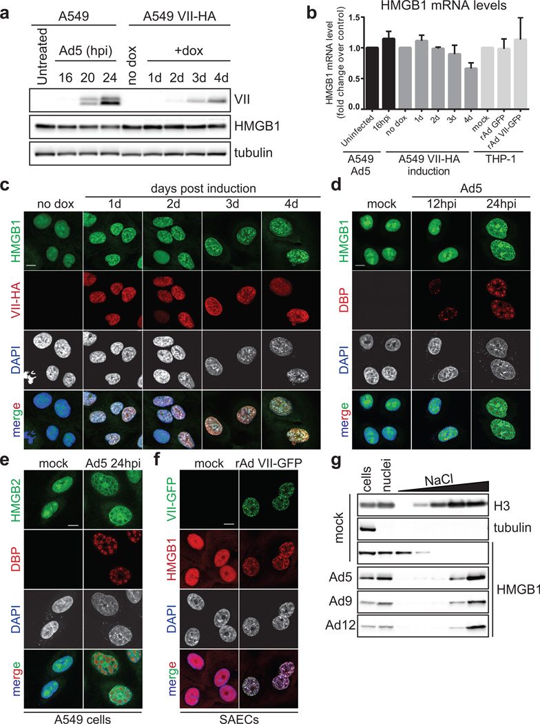
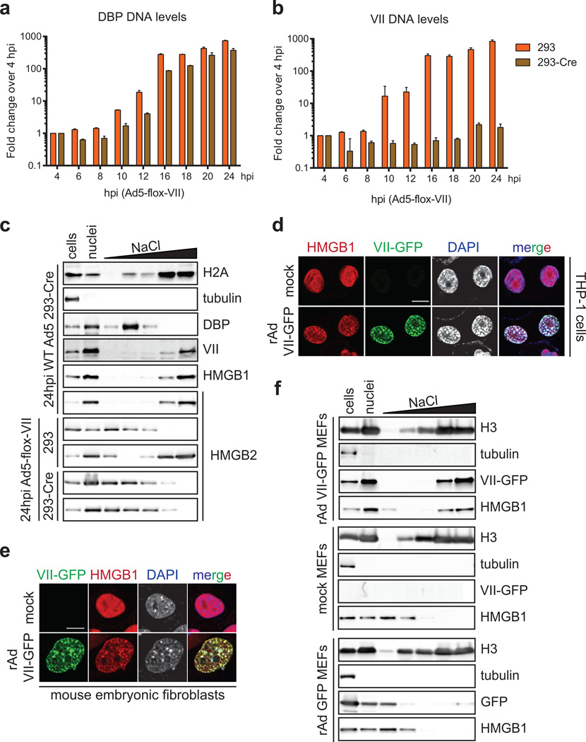
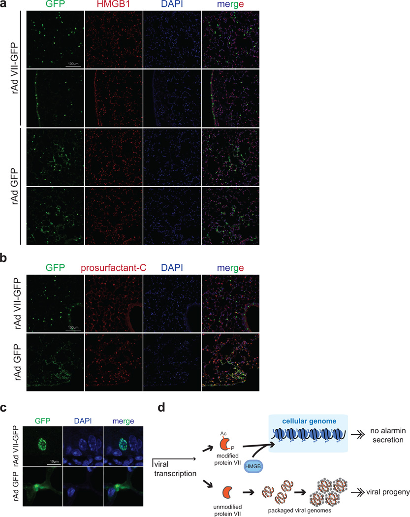
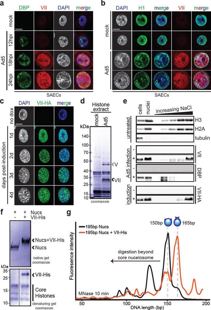
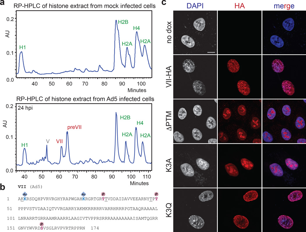
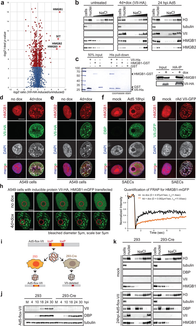
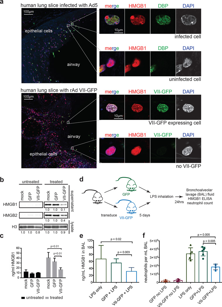
Viral proteins mimic host protein structure and function to redirect cellular processes and subvert innate defenses. Small basic proteins compact and regulate both viral and cellular DNA genomes. Nucleosomes are the repeating units of cellular chromatin and play an important part in innate immune responses. Viral-encoded core basic proteins compact viral genomes, but their impact on host chromatin structure and function remains unexplored. Adenoviruses encode a highly basic protein called protein VII that resembles cellular histones. Although protein VII binds viral DNA and is incorporated with viral genomes into virus particles, it is unknown whether protein VII affects cellular chromatin. Here we show that protein VII alters cellular chromatin, leading us to hypothesize that this has an impact on antiviral responses during adenovirus infection in human cells. We find that protein VII forms complexes with nucleosomes and limits DNA accessibility. We identified post-translational modifications on protein VII that are responsible for chromatin localization. Furthermore, proteomic analysis demonstrated that protein VII is sufficient to alter the protein composition of host chromatin. We found that protein VII is necessary and sufficient for retention in the chromatin of members of the high-mobility-group protein B family (HMGB1, HMGB2 and HMGB3). HMGB1 is actively released in response to inflammatory stimuli and functions as a danger signal to activate immune responses. We showed that protein VII can directly bind HMGB1 in vitro and further demonstrated that protein VII expression in mouse lungs is sufficient to decrease inflammation-induced HMGB1 content and neutrophil recruitment in the bronchoalveolar lavage fluid. Together, our in vitro and in vivo results show that protein VII sequesters HMGB1 and can prevent its release. This study uncovers a viral strategy in which nucleosome binding is exploited to control extracellular immune signaling.


Figures

Extended Data Figure 1
Extended Data Figure 1. Adenovirus protein VII distorts chromatin
a, Protein VII localizes to cellular chromatin and viral replication centers in U2OS similarly to SAECs in Fig 1a. b, Protein VII mRNA levels measured by quantitative PCR showing that after 4 days of induction in the A549 cell line, the level of protein VII transcripts is approximately 10% of that measured during infection at 16 hpi. Despite the low relative level, this amount of protein VII is sufficient to cause dramatic changes in the nucleus (error bars=±s.d., nbiological=3). c, Inducible cell lines of U2OS and HeLa expressing protein VII-HA show chromatin localization and distortion, similar to A549 cells in Fig 1c. d, Inducible A549 cell lines expressing viral protein V, the precursor for protein VII (preVII) or cellular protamine PRM1 with C-terminal HA tags. Although all 3 proteins possess a large number of charged residues, none are sufficient to distort cellular chromatin or increase nuclear size as observed with mature protein VII.
Extended Data Figure 2
Extended Data Figure 2. Protein VII associates tightly with chromatin and binds DNA and nucleosomes in vitro
a, Western blot analysis showing protein VII in histone extracts from infected HeLa cells at 24 hpi. b, Chromatin fractionation of lysates from A549 cells that were uninfected (mock) or infected for 24 hrs with Ad5. Viral and cellular proteins were detected by western blotting with various antibodies as indicated. c, Agarose gel analysis of DNA extracted from nuclear fractionation experiments indicating the size of DNA is between 100 – 200 bp and elutes predominantly in the higher salt fractions. d, Chromatin fractionation of cells induced to express protein VII indicating protein VII present in the highest salt fraction from the first day of induction. e–f, Recombinant protein VII-His binds DNA. Incubating increasing molar amounts of protein VII with 195bp DNA results in shifts by native gel electrophoresis indicating protein VII-DNA complex formation. Staining with either ethidium bromide (e) or coomassie (f) are shown to verify the presence of DNA and protein, respectively. g, Ethidium bromide staining shows DNA content of nucleosome shifts from gel in Fig 1f.
Extended Data Figure 3
Extended Data Figure 3. Bioanalyzer examination of MNase digested nucleosomes and protein VII-nucleosome complexes
a, 195bp Nucleosomes or protein VII-nucleosome complexes were incubated with MNase for the indicated times, the reaction was stopped, DNA extracted and analyzed. As in Fig 1g, nucleosomes are shown in black and protein VII-nucleosome complexes in orange. The presence of protein VII pauses digestion at 165bp, suggesting that protein VII is blocking access to the DNA. b, 147bp nucleosomes or protein VII-nucleosome complexes were incubated with MNase for the indicated times, the reaction was stopped, DNA extracted and analyzed. Graphs show nucleosomes in grey and protein VII-nucleosome complexes in orange. The presence of protein VII completely blocks digestion even after nucleosomes alone have been digested well beyond the core particle. In contrast to what would be expected for linker histones, protein VII protects the core nucleosome particle from digestion. These data indicate that protein VII may be masking the substrate for MNase through complex formation. This represents a unique mechanism of nucleosome binding and suggests a model for blocking DNA access in cellular chromatin during infection.
Extended Data Figure 4
Extended Data Figure 4. Purification of protein VII from infected cells
a, Coomassie stained SDS-PAGE analysis of fractions from RP-HPLC in Fig 2a. The bands in fraction 38–41 min correspond to histone H1. Protein VII and V, as indicated, were verified by mass spectrometry analysis (not shown). The slight upward shift of the protein VII bands in the later peak corresponds to the higher abundance of protein preVII, as seen by HPLC in Fig 2a. b, Western blot analysis of protein VII in HPLC fractions from (a). c, Time-course of infection followed by histone extraction and HPLC analysis. Mass spectrometry analysis verified peaks in each sample as indicated.
Extended Data Figure 5
Extended Data Figure 5. Representative mass spectra
Annotated MS/MS spectra of identified peptides of protein VII containing PTMs (a–c, acetylated peptides; d–f, phosphorylated peptides). The images represent the observed fragment ions collected using MS/MS collision induced dissociation (CID). Colored lines represent matches between observed and expected fragment ions of the given peptides. Specifically, green lines represent not fragmented precursor mass, blue lines represent matches with y-type fragments, red lines with b-type fragments, and yellow boxed masses represent fragments containing PTM neutral losses (e.g. ions that lost the phosphorylation during fragmentation).
Extended Data Figure 6
Extended Data Figure 6. Acetylated VII spectra from virus particles and analysis of total histone PTM changes upon protein VII expression
a, LC-MS analysis of unmodified and modified chymotryptic peptide AKKRSDQHPVRVRGHY. On the left, nanoLC–MS extracted ion chromatograms of protein VII peptides identified in the histone extracts of adenovirus infected cells (Inf) or viral particles (VP). The top left represents the modified form, while the bottom left the unmodified form. Non-modified forms were detected in both conditions for VII and VP, while the acetylated form was unique for the infected sample only (Inf). On the right, full MS spectrum of the modified (top) and unmodified (bottom) peptide. Circled mass represents the monoisotopic signal of the peptide. b, Summary of post-translational modifications detected on protein VII. Peptides shown were identified during infection at various time points with the mature protein VII in the top row and the precursor, preVII, in the bottom row. The numbers in brackets for preVII indicate the location of the same moiety in mature protein VII. Acetylation sites were detected in approximately 3% of peptides for mature protein VII and 2% of peptides in preVII. Phosphorylation was detected in approximately 1% of peptides for mature protein VII and preVII. c–d, Quantification of histone H3 (c) and H4 (d) PTMs in protein VII-HA induced (+dox) and uninduced (−dox) A549 cells from the analysis of crude histone mixtures (nbiological=3). Positions of PTMs are listed along the x-axis. Modification type is indicated by color as shown. y-axis represents the cumulative extent of PTMs as relative to the total histone H3 or H4, respectively. e, Breakdown of the histone marks (H3K14ac, H3K27me1, H3K36me3, H4K20me1, H4K20me2, and H4K20me3) found significantly different (nbiological=3) in terms of relative abundance between the protein VII-HA induced and uninduced states (<5% homoscedastic two-tailed t-test). Error bars represent ± standard deviation.
Extended Data Figure 7
Extended Data Figure 7. Bioinformatic analysis of proteins enriched in the high salt fraction upon protein VII expression
a, Venn diagram showing overlap between three biological replicates of high salt fraction proteins significantly enriched as compared to uninduced cells. b, Proteins found significantly enriched in the protein VII-HA induced state as compared to uninduced (<5% homoscedastic t-test) in all three biological replicates (VII-HA induced: proteins identified only in protein VII-HA induced condition). c–d, Classification of proteins significantly enriched in minimum two out of three biological replicates (protein VII-HA induced vs uninduced) according to process network enrichment and gene ontology (GO) biological process (GeneGo's MetaCore pathways analysis package; false discovery rate <5%); each GO term was ranked using p-value enrichment.
Extended Data Figure 8
Extended Data Figure 8. Protein VII retains HMGB1 and HMGB2 in chromatin
a, Western blot of adenovirus infected or doxycycline treated A549 cells showing the relative levels of protein VII expression. HMGB1 levels do not change upon infection or protein VII expression. Tubulin is shown as a loading control. b, Quantitative PCR analysis of mRNA transcripts of HMGB1 in various cell types as indicated (for A549, nbiological=3, for THP-1, nbiological=2, error bar=±s.d.). The levels of HMGB1 do not significantly change. c, Immunofluorescence analysis of a time-course of protein VII-HA (red) induction shown with HMGB1 (green) and DAPI (grey, blue in merge) in A549 cells. Expression of protein VII-HA results in a change to the HMGB1 distribution upon expression. d, HMGB1 (green) localization changes between 12 and 24 hpi of wild-type adenovirus in A549 cells, and adopts a pattern similar to protein VII as in Fig 1a. DBP (red) is shown as a marker of infection, DNA is stained with DAPI (blue in merge). e, Same as (d) showing HMGB2 adopts the same pattern as HMGB1 during Ad5 infection at 24 hpi. f, Multiple cells showing the same pattern of HMGB1 re-localization upon expressing VII-GFP as in Fig 3g. g, HMGB1 retention in the high salt fraction is conserved across Ad serotypes. Western blot analysis of HMGB1 from salt fractionated A549 cells infected with Ad5, Ad9 or Ad12 as shown.
Extended Data Figure 9
Extended Data Figure 9. Protein VII is necessary and sufficient for chromatin retention of HMGB1 in human and mouse cells
a–b, Replication of Ad5-flox-VII virus on 293 or 293-Cre cells. Quantitative PCR analysis of viral genomic DNA over a time-course of infection (a) shows the DBP gene is increasing exponentially in 293 and 293-Cre cells when infected with Ad5-flox-VII virus. In contrast, PCR for the protein VII gene (b) demonstrates deletion in 293-Cre cells (nbiological=2, error bar=±s.d.). c, Salt fractionation of 293-Cre cells infected with wild-type Ad5 indicating that the Cre recombinase does not interfere with the ability of protein VII to retain HMGB1 in the high salt chromatin fraction. Protein VII is also necessary for the chromatin retention of HMGB2. d, THP-1 cells transduced to express protein VII-GFP results in chromatin distortion and HMGB1 retention in chromatin. Immunofluorescence of transduced PMA-treated THP-1 cells showing protein VII-GFP (green), HMGB1 (red) and DNA (grey, blue in merge). e, Transduction to express protein VII-GFP is sufficient to relocalize mouse HMGB1 in mouse embryonic fibroblast (MEF) cells. f, Salt fractionation of mouse embryonic fibroblast cells transduced to express protein VII-GFP. Human Ad5 protein VII is sufficient to retain mouse HMGB1 in the high salt fraction in MEF cells. The control vector expressing GFP alone does not have this effect.
Extended Data Figure 10
Extended Data Figure 10. Transduction of mouse lungs demonstrating expression of GFP or protein VII-GFP
a, Sections of mouse lungs transduced to express protein VII-GFP or GFP co-stained for HMGB1. GFP signal shows multiple cell types transduced in both cases. Protein VII-GFP has a more distinct nuclear signal than GFP, which also appears cytoplasmic. Two sections for each condition are shown to indicate transduction efficiency. b, Same as (a) but co-stained for prosurfactant-C to mark type II pneumocytes. Some cells are positive for both, confirming multiple cell types transduced. c, Zoomed images of individual epithelial cells from mouse lungs showing the characteristic protein VII-GFP pattern colocalizing with DAPI in the nucleus. GFP only is mostly cytoplasmic. d, Schematic summarizing function of protein VII during infection. Newly synthesized protein VII late during infection can be post-translationally modified and binds to HMGB1, sequestering it on the cellular chromatin and preventing its release. Unmodified protein VII is packaged in viral progeny.
Figure 1
Figure 1. Protein VII is sufficient to alter chromatin and directly binds nucleosomes
a–b Ad5-infected SAECs stained for protein VII (red) with DBP (a), or histone H1 (b), and DAPI (grey, blue in merge). c, Protein VII-HA induced cells over four days showing HA (green) and DAPI (grey, blue in merge). d, SDS-PAGE of histone extract from Ad5-infected cells showing protein V and protein VII. e, Western blot of chromatin fractionation from nuclei of Ad5-infected cells, induced for protein VII-HA, or untreated. f, Protein VII binds to nucleosomes. Protein bands from native gel stained with coomassie (top) were subjected to 2D analysis by SDS-PAGE (bottom). g, Protein VII protects nucleosome complexes from MNase digestion. Bioanalyzer curves represent nucleosomes alone (black) or protein VII-nucleosome complexes (orange).
Figure 2
Figure 2. Post-translational modifications on protein VII contribute to chromatin localization
a, RP-HPLC analysis of histone extracts. Viral proteins V, VII and preVII are indicated at 24 hpi. b, Primary sequence of protein VII with modified residues identified in infected cells. Underlined residues represent moieties that may also be modified in identified peptides (see ED). c, Immunofluoresence showing DAPI (grey, blue in merge) and protein VII (red) as wild-type or with alanine substitutions at PTM sites (ΔPTM), K3A or K3Q.
Figure 3
Figure 3. Protein VII directly binds HMGB1 and is necessary for retention of the alarmin in cellular chromatin
a, Volcano plot for proteomics analysis of one representative biological replicate of high salt fraction. The y-axis represents −log2 statistical p-value and x-axis represents log2 protein fold-change between uninduced or protein VII expressing cells (homoscedastic two-tailed t-test, p<0.05 red dots; ntechnical=3). b, Nuclear fractionation shows HMGB1 and HMGB2 normally elute from nuclei at low salt concentrations but are retained in high salt fractions by protein VII-HA. c, Protein VII interacts with HMGB1 in pull-down of recombinant HMGB1-GST (left, coomassie-stained SDS-PAGE) and immunoprecipitation of HMGB1 (right, western blots). d–e, Protein VII expression alters localization of HMGB1 (d) and HMGB2 (e). Immunofluorescence shows protein VII-HA (green) co-localized with HMGB1 (d) and HMGB2 (e) in cellular chromatin, DAPI (grey, blue in merge). f, Same as (d) at 18 hpi with Ad5 DBP (green). g, Protein VII-GFP relocalizes HMGB1 (red) to chromatin with DAPI (grey, blue in merge). h, FRAP experiment with HMGB1-mGFP. Recovery of FRAP signal in time-course images (left) with quantification and diffusion coefficients (right). i, Schematic showing loxP strategy for deleting protein VII. j, Western blots comparing 293 and 293Cre cells infected with Ad5-flox-VII virus. k, Salt fractionation in nuclei from (j).
Figure 4
Figure 4. Protein VII prevents HMGB1 release
a, Precision cut lung slices (PCLS) infected with Ad5 or transduced to express protein VII-GFP. Endogenous HMGB1 (red) is redistributed in cells with virus (DBP in top) and VII-GFP (bottom). b, Protein VII-GFP is sufficient to inhibit HMGB1 and HMGB2 release in THP-1 cells. Numbers indicate relative intensities of bands quantified with ImageJ. c, ELISA-based quantification of HMGB1 in supernatants from (b), error bars=±s.d., ntechnical=4, homoscedastic one-tailed t-test. d, Schematic for investigating protein VII in a mouse lung injury model. e, Expression of protein VII-GFP decreases HMGB1 in mouse BAL fluid as quantified by ELISA, error bars=±s.d., biological replicates: nLPS=4, nGFP+LPS=6, nVII-GFP+LPS=7, homoscedastic one-tailed (p=0.02) or two-tailed (p=0.003) t-test. f, Neutrophils in BAL fluid are significantly fewer in mice expressing protein VII-GFP, error bars=±s.d., biological replicates: nGFP+LPS=6, nVII-GFP+LPS=4, nLPS=5, nGFP=3, nVII-GFP=3 homoscedastic two-tailed t-test.

Comment in

Similar articles

Cited by

References

    1. Elde NC, Malik HS. The evolutionary conundrum of pathogen mimicry. Nature Reviews Microbiology. 2009;7:787–797. - PubMed
    1. Smale ST, Tarakhovsky A, Natoli G. Chromatin contributions to the regulation of innate immunity. Annu. Rev. Immunol. 2014;32:489–511. - PubMed
    1. Lischwe MA, Sung MT. A histone-like protein from adenovirus chromatin. Nature. 1977;267:552–554. - PubMed
    1. Chatterjee PK, Vayda ME, Flint SJ. Adenoviral protein VII packages intracellular viral DNA throughout the early phase of infection. EMBO J. 1986;5:1633–1644. - PMC - PubMed
    1. Vayda ME, Rogers AE, Flint SJ. The structure of nucleoprotein cores released from adenovirions. Nucleic Acids Research. 1983;11:441–460. - PMC - PubMed

Methods References

    1. Khandelia P, Yap K, Makeyev EV. Streamlined platform for short hairpin RNA interference and transgenesis in cultured mammalian cells. Proc. Natl. Acad. Sci. U.S.A. 2011;108:12799–12804. - PMC - PubMed
    1. Kozarsky KF, Jooss K, Donahee M, Strauss JF, Wilson JM. Effective treatment of familial hypercholesterolaemia in the mouse model using adenovirus-mediated transfer of the VLDL receptor gene. Nat. Genet. 1996;13:54–62. - PubMed
    1. Orazio NI, Naeger CM, Karlseder J, Weitzman MD. The adenovirus E1b55K/E4orf6 complex induces degradation of the Bloom helicase during infection. Journal of Virology. 2011;85:1887–1892. - PMC - PubMed
    1. Le LP, et al. Core labeling of adenovirus with EGFP. Virology. 2006;351:291–302. - PMC - PubMed
    1. Reich NC, Sarnow P, Duprey E, Levine AJ. Monoclonal antibodies which recognize native and denatured forms of the adenovirus DNA-binding protein. Virology. 1983;128:480–484. - PubMed

Publication types

MeSH terms