. 2016 Jun 30;12(6):e1006146.
doi: 10.1371/journal.pgen.1006146. eCollection 2016 Jun.

The Meiotic Recombination Activator PRDM9 Trimethylates Both H3K36 and H3K4 at Recombination Hotspots In Vivo

Affiliations

The Meiotic Recombination Activator PRDM9 Trimethylates Both H3K36 and H3K4 at Recombination Hotspots In Vivo

Natalie R Powers et al. PLoS Genet. .

Abstract

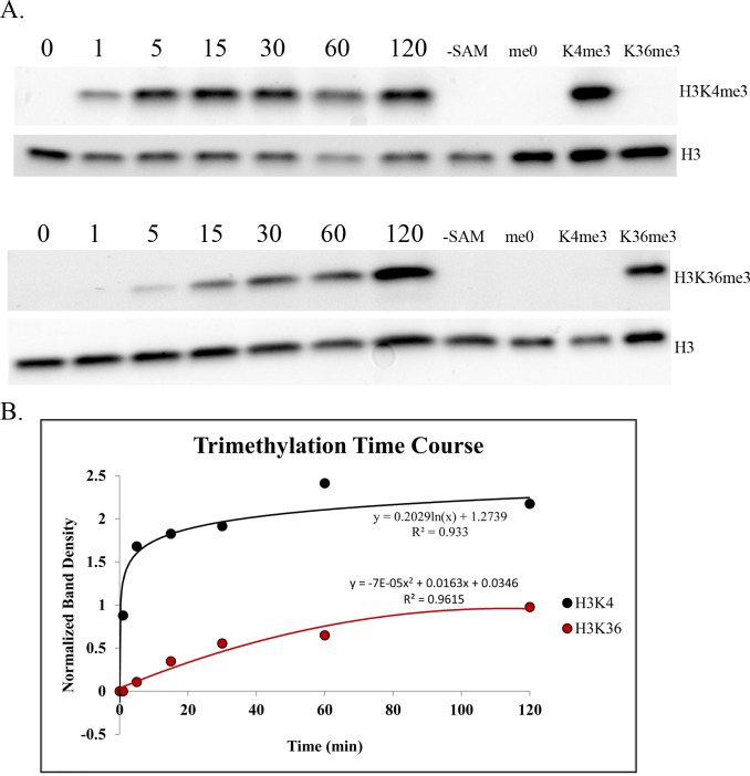
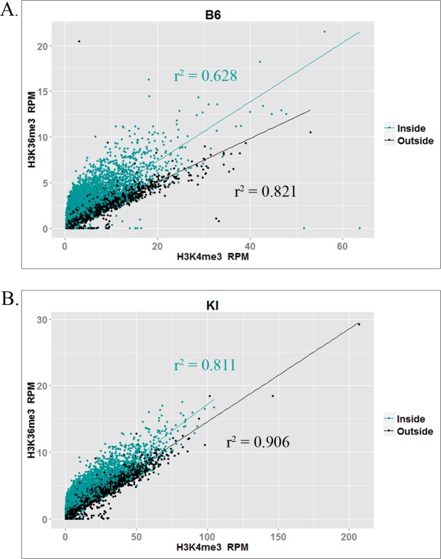
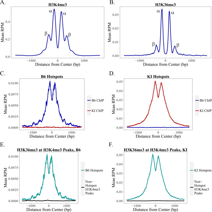
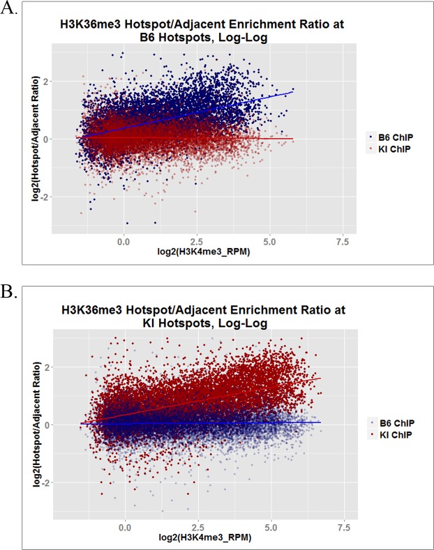
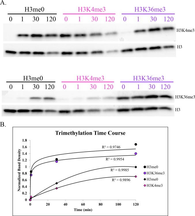
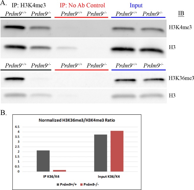
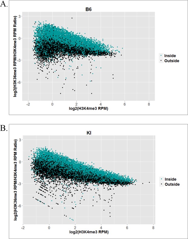
In many mammals, including humans and mice, the zinc finger histone methyltransferase PRDM9 performs the first step in meiotic recombination by specifying the locations of hotspots, the sites of genetic recombination. PRDM9 binds to DNA at hotspots through its zinc finger domain and activates recombination by trimethylating histone H3K4 on adjacent nucleosomes through its PR/SET domain. Recently, the isolated PR/SET domain of PRDM9 was shown capable of also trimethylating H3K36 in vitro, raising the question of whether this reaction occurs in vivo during meiosis, and if so, what its function might be. Here, we show that full-length PRDM9 does trimethylate H3K36 in vivo in mouse spermatocytes. Levels of H3K4me3 and H3K36me3 are highly correlated at hotspots, but mutually exclusive elsewhere. In vitro, we find that although PRDM9 trimethylates H3K36 much more slowly than it does H3K4, PRDM9 is capable of placing both marks on the same histone molecules. In accord with these results, we also show that PRDM9 can trimethylate both K4 and K36 on the same nucleosomes in vivo, but the ratio of K4me3/K36me3 is much higher for the pair of nucleosomes adjacent to the PRDM9 binding site compared to the next pair further away. Importantly, H3K4me3/H3K36me3-double-positive nucleosomes occur only in regions of recombination: hotspots and the pseudoautosomal (PAR) region of the sex chromosomes. These double-positive nucleosomes are dramatically reduced when PRDM9 is absent, showing that this signature is PRDM9-dependent at hotspots; the residual double-positive nucleosomes most likely come from the PRDM9-independent PAR. These results, together with the fact that PRDM9 is the only known mammalian histone methyltransferase with both H3K4 and H3K36 trimethylation activity, suggest that trimethylation of H3K36 plays an important role in the recombination process. Given the known requirement of H3K36me3 for double strand break repair by homologous recombination in somatic cells, we suggest that it may play the same role in meiosis.


Conflict of interest statement

The authors have declared that no competing interests exist.

Figures

Fig 1
Fig 1. Intrinsic H3K4 and H3K36 trimethylation activities of the PRDM9 PR/SET domain.
(A) Western blots of a typical histone methyltransferase assay time course for PRDM9 PR/SET domain, with unmodified recombinant histone H3 as substrate. Duplicate blots were probed with α-H3K4me3 and α-H3K36me3 antibody, respectively. Both blots were then stripped and re-probed with α-H3 antibody to determine the total amount of histone H3 in each lane. Numbers indicate time in minutes; “-SAM” indicates a negative control reaction that was run without the methyl donor S-adenosylmethionine; the last three lanes contain the denoted recombinant histone, to demonstrate antibody specificity. (B) Histone methyltransferase time course plot. The y-axis represents the ratio of the density of each H3K4me3 or H3K36me3 band to the corresponding H3 band, to control for the total amount of histone H3 in each lane.
Fig 2
Fig 2. Correlation between H3K4me3 and H3K36me3 at hotspots.
Plots show correlations between normalized (RPM) H3K4me3 and H3K36me3 ChIP-seq reads at individual hotspots in (A) the B6 strain and (B) the KI strain. Trendlines and correlation coefficients are shown for the subset of hotspots inside (teal) and outside (black) transcribed genes.
Fig 3
Fig 3. Aggregation Plots for H3K4me3 and H3K36me3 at Hotspots.
(A-B) These aggregation plots show average H3K4me3 and H3K36me3 enrichment at 12,774 B6 hotspots for which we have previously determined the location of the 36bp PRDM9 binding site with high confidence. These plots, centered at the PRDM9 binding site, show the shape of the average H3K4me3 and H3K36me3 peak at a hotspot. (C-D) show average H3K36me3 enrichment across B6 (C) and KI (D) hotspots, using ChIP-seq data from the B6 and KI strains. Like PRDM9-dependent H3K4me3, H3K36me3 enrichment at hotspots is strain-specific. (E-F) show average H3K36me3 enrichment in hotspots specific to the denoted strain, and at non-hotspot H3K4me3 peaks (e.g. promoters). As measured by ChIP-seq, H3K4me3 and H3K36me3 coincide only at hotspots.
Fig 4
Fig 4. H3K36me3 inside and outside hotspots in actively transcribed genes.
These log-log plots show, for hotspots located within actively transcribed genes, the ratio of normalized H3K36me3 ChIP-seq enrichment (RPM) within each hotspot to that within a same-sized region immediately adjacent to each hotspot, as a function of PRDM9-dependent H3K4me3 enrichment at each hotspot. (A) shows the data from both strains at B6 hotspots, while (B) shows the data from both strains at KI hotspots.
Fig 5
Fig 5. Intrinsic ability of PRDM9 PR/SET domain to trimethylate H3K4 and H3K36 on histones previously trimethylated for the other mark.
(A) Western blots of a single histone methyltransferase assay time course, measuring the H3K4 and H3K36 trimethylation activity of recombinant PRDM9 PR/SET domain, using recombinant unmodified histone H3 (black) H3K4me3 (pink) or H3K36me3 (purple) as substrate. Duplicate blots were probed with α-H3K4me3 and α-H3K36me3 antibodies, respectively. Both blots were then stripped and re-probed with α-Histone H3 antibody. Numbers indicate time in minutes. (B) Methyltransfease assay time course, quantified by densitometry and normalized to histone H3 as in Fig 1. Circles represent H3K4 trimethylation, while diamonds represent H3K36 trimethylation, on the histones denoted in the legend.
Fig 6
Fig 6. Detection of H3K4me3/H3K36me3-double-positive nucleosomes in vivo by immunoprecipitation.
(A) This figure shows immunoprecipitation and immunoblotting of histones extracted from 14dpp mouse spermatocytes, from either C57BL/6J mice (Prdm9+/+) or homozygous B6;129P2-Prdm9tm1Ymat/J mice (Prdm9-/-). Immunoprecipitation was done using 4μg acid-extracted histone, with either α-H3K4me3 antibody or no antibody as a control. Input samples were prepared with 0.5μg acid-extracted histone. Equal amounts of each IP and Input were run on two identical gels, which were transferred to nitrocellulose and immunoblotted with either α-H3K4me3 or α-H3K36me3 antibody. Both blots were then stripped and re-probed with α-Histone H3 antibody to confirm equal loading of total histone. See S5 Fig for a biological replicate with an additional re-probe for histone H4. (B) This chart shows the ratios of H3K36me3 signal to H3K4me3 signal, as quantified by densitometry, in the Prdm9+/+ and Prdm9-/- IPs and inputs. The H3K4me3 and H3K36me3 bands for both the IPs and inputs were normalized to the histone H3 band in the corresponding input.
Fig 7
Fig 7. Log-log plots showing the ratio of H3K36me3 to H3K4me3 at hotspots as a function of PRDM9-dependent H3K4me3 enrichment, inside and outside actively transcribed genes.
(A) shows the B6 hotspot data in the B6 strain; (B) shows the KI hotspot data in the KI strain.

References

    1. Chan AH, Jenkins PA, Song YS (2012) Genome-wide fine-scale recombination rate variation in Drosophila melanogaster. PLoS Genet 8: e1003090 10.1371/journal.pgen.1003090 - DOI - PMC - PubMed
    1. Kaur T, Rockman MV (2014) Crossover heterogeneity in the absence of hotspots in Caenorhabditis elegans. Genetics 196: 137–148. 10.1534/genetics.113.158857 - DOI - PMC - PubMed
    1. Tischfield SE, Keeney S (2012) Scale matters: the spatial correlation of yeast meiotic DNA breaks with histone H3 trimethylation is driven largely by independent colocalization at promoters. Cell Cycle 11: 1496–1503. 10.4161/cc.19733 - DOI - PMC - PubMed
    1. Choi K, Zhao X, Kelly KA, Venn O, Higgins JD, et al. (2013) Arabidopsis meiotic crossover hot spots overlap with H2A.Z nucleosomes at gene promoters. Nat Genet 45: 1327–1336. 10.1038/ng.2766 - DOI - PMC - PubMed
    1. Parvanov ED, Petkov PM, Paigen K (2010) Prdm9 controls activation of mammalian recombination hotspots. Science 327: 835 10.1126/science.1181495 - DOI - PMC - PubMed

Publication types