. 2016 Jun 30;11(6):e0157231.
doi: 10.1371/journal.pone.0157231. eCollection 2016.

Rickettsia australis Activates Inflammasome in Human and Murine Macrophages

Affiliations

Rickettsia australis Activates Inflammasome in Human and Murine Macrophages

Claire Smalley et al. PLoS One. .

Abstract

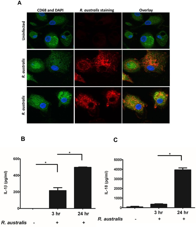
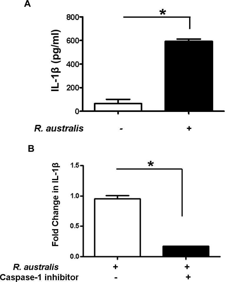
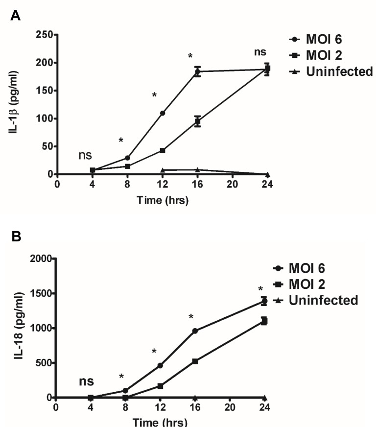
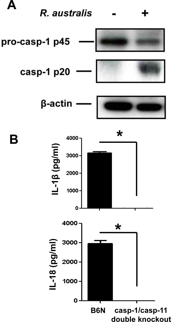
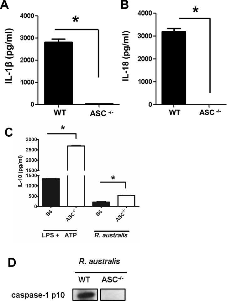
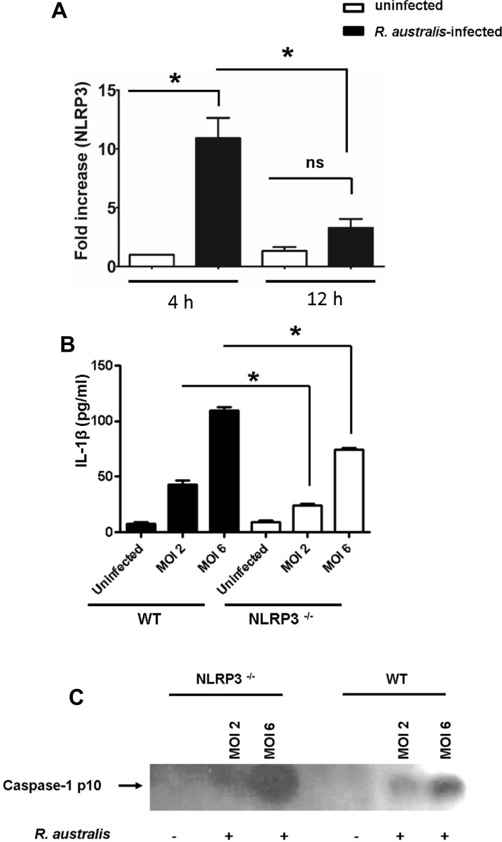
Rickettsiae actively escape from vacuoles and replicate free in the cytoplasm of host cells, where inflammasomes survey the invading pathogens. In the present study, we investigated the interactions of Rickettsia australis with the inflammasome in both mouse and human macrophages. R. australis induced a significant level of IL-1β secretion by human macrophages, which was significantly reduced upon treatment with an inhibitor of caspase-1 compared to untreated controls, suggesting caspase-1-dependent inflammasome activation. Rickettsia induced significant secretion of IL-1β and IL-18 in vitro by infected mouse bone marrow-derived macrophages (BMMs) as early as 8-12 h post infection (p.i.) in a dose-dependent manner. Secretion of these cytokines was accompanied by cleavage of caspase-1 and was completely abrogated in BMMs deficient in caspase-1/caspase-11 or apoptosis-associated speck-like protein containing a caspase activation and recruitment domain (ASC), suggesting that R. australis activate the ASC-dependent inflammasome. Interestingly, in response to the same quantity of rickettsiae, NLRP3-/- BMMs significantly reduced the secretion level of IL-1β compared to wild type (WT) controls, suggesting that NLRP3 inflammasome contributes to cytosolic recognition of R. australis in vitro. Rickettsial load in spleen, but not liver and lung, of R. australis-infected NLRP3-/- mice was significantly greater compared to WT mice. These data suggest that NLRP3 inflammasome plays a role in host control of bacteria in vivo in a tissue-specific manner. Taken together, our data, for the first time, illustrate the activation of ASC-dependent inflammasome by R. australis in macrophages in which NLRP3 is involved.


Conflict of interest statement

Competing Interests: The authors have declared that no competing interests exist.

Figures

Fig 1
Fig 1. Infection of human PBMC-derived macrophages with R. australis and activation of inflammasome.
Human PBMC-derived macrophages were prepared and infected with R. australis. Cells and culture supernatant were collected at 24 h p.i.. A, Cytosolic rickettsiae were detected by confocal immunofluorescence microscopy after infection with R. australis at an MOI of 5. Images were acquired using 60 × magnification with a water immersion lens. Macrophages are depicted in green, nuclei (DAPI) in blue, and R. australis in red. Secretion levels of IL-1β (B) and IL-18 (C) at 3 h and 24 h p.i. by human PBMC-derived macrophages infected with R. australis at an MOI of 2 were determined by ELISA. Data represent two independent experiments with consistent results. Each experiment included at least 4 replicates. *, p<0.05.
Fig 2
Fig 2. Activation of inflammasome by rickettsiae in THP-1 derived macrophages and caspase-1-dependent secretion of IL-1β.
Human THP-1 cells were differentiated to macrophages using PMA, and infected with R. australis at an MOI of 5. A, Infection with R. australis induced a significant increase in IL-1β production compared to uninfected cells at 24 h p.i.. B, Inhibition of caspase-1 significantly reduced the secretion levels of IL-1β by R. autralis-infected THP-1 derived macrophages. The fold change in IL-1β by treated cells vs. untreated controls was calculated and compared. Data represent two independent experiments with consistent results. Each experiment included at least 4 replicates. *, p<0.05.
Fig 3
Fig 3. Kinetics and dose-dependent mechanisms of secretion of IL-1β and IL-18 by Rickettsia-infected BMMs.
WT BMMs were isolated, cultivated, and infected with R. australis at MOI of 2 or 6. Cell culture supernatants were harvested at 4 hour intervals over 24 h p.i.. Secretion of IL-1β (A) and IL-18 (B) was determined by ELISA. Data represent two independent experiments with consistent results. Each experiment included at least 4 replicates. *, p<0.05; ns, not significantly different.
Fig 4
Fig 4. R. australis activated inflammasome in BMMs.
A, WT BMMs were isolated, cultivated and infected with R. australis at an MOI of 10. At 24 h p.i., culture supernatant and cell lysates were collected. Cell lysates were processed for detection of activation of caspase-1. B, BMMs of B6N and caspase-1/11-double knockout mice were isolated and infected with R. australis as described above. The secretion levels of IL-1β and IL-18 were determined by ELISA. *, p<0.05.
Fig 5
Fig 5. ASC-dependent inflammasome recognized cytosolic rickettsiae.
BMMs of WT and ASC-/- mice were isolated, cultivated, and infected with R. australis at an MOI of 10 or treated with LPS plus ATP as described in Materials and Methods. At 24 h p.i., the secretion of IL-1β (A), IL-18 (B) and IL-10 (C) was assayed by ELISA. D, Activation of caspase-1 was determined by immunoblotting detection of the active unit p10 in the processed supernatant of infected WT and ASC-/- BMMs at 24 h p.i.. Data represent two independent experiments with consistent results. Each experiment included at least 4 replicates. *, p<0.05.
Fig 6
Fig 6. NLRP3 was involved in recognition of rickettsiae by inflammasome at the early stage of infection in BMMs.
WT and NLRP3-/- BMMs were isolated, cultivated, and infected with R. australis at MOIs of 6 and 2. The transcriptional levels of NLRP3 in WT BMMs at different time intervals of infection (at MOI of 6) were determined by RT-PCR as described in Materials and Methods (A). At 12 h (B) p.i., the secretion levels of IL-1β by WT and NLRP3-/- BMMs were determined by ELISA. The cleavage of caspase-1 was determined by detection of the active unit p10 in the processed supernatant at 12 h p.i. (C). Data represent mean ± SD for at least 3 replicates each group. *, p<0.05 for a significant difference between WT and NLRP3-/- mice; ns, not significantly different.

References

    1. Walker DH. Rickettsiae and rickettsial infections: the current state of knowledge. Clin Infect Dis. 2007; 45 Suppl 1:S39–44. - PubMed
    1. Schroder K, Tschopp J. The inflammasomes. Cell. 2010;140(6):821–32. 10.1016/j.cell.2010.01.040 - DOI - PubMed
    1. Casson CN, Shin S. Inflammasome-mediated cell death in response to bacterial pathogens that access the host cell cytosol: lessons from Legionella pneumophila. Front Cell Infect Microbiol. 2013; 3:111 10.3389/fcimb.2013.00111 - DOI - PMC - PubMed
    1. Li H, Willingham SB, Ting JP, Re F. Cutting edge: inflammasome activation by alum and alum's adjuvant effect are mediated by NLRP3. J Immunol. 2008; 181(1):17–21. - PMC - PubMed
    1. Feng HM, Popov VL, Walker DH. Depletion of gamma interferon and tumor necrosis factor alpha in mice with Rickettsia conorii-infected endothelium: impairment of rickettsicidal nitric oxide production resulting in fatal, overwhelming rickettsial disease. Infect Immun. 1994; 62(5):1952–60. - PMC - PubMed

LinkOut - more resources