

Existence of Normal Limbal Epithelium in Eyes With Clinical Signs of Total Limbal Stem Cell Deficiency
- PMID: 27362882
- PMCID: PMC5067954
- DOI: 10.1097/ICO.0000000000000914
Existence of Normal Limbal Epithelium in Eyes With Clinical Signs of Total Limbal Stem Cell Deficiency
Abstract
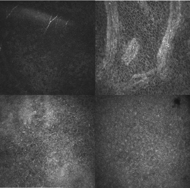
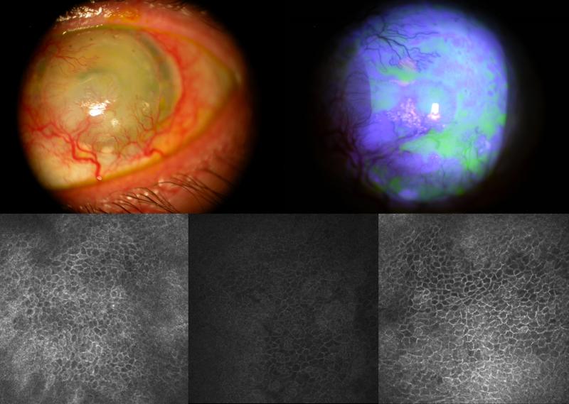
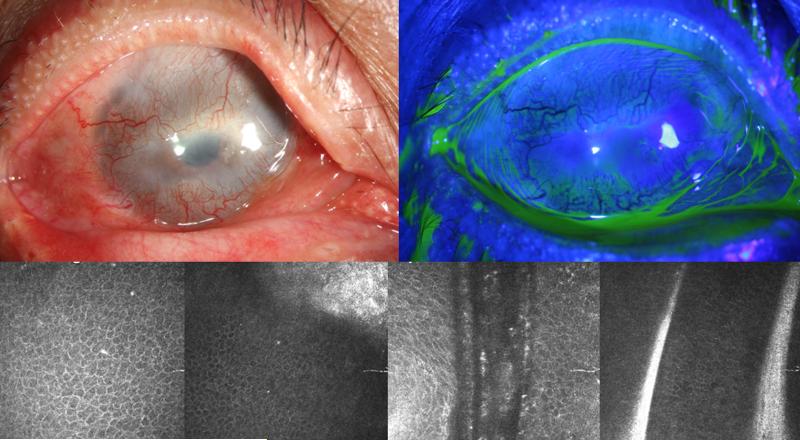
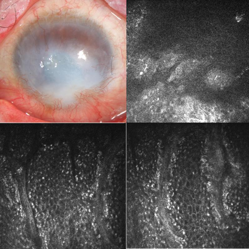
Purpose: To report the presence of normal limbal epithelium detected by in vivo confocal laser scanning microscopy (IVCM) in 3 cases of clinically diagnosed total limbal stem cell deficiency (LSCD).
Methods: This is a retrospective case report consisting of 3 patients who were diagnosed with total LSCD based on clinical examination and/or impression cytology. Clinical data including ocular history, presentation, slit-lamp examination, IVCM, and impression cytology were reviewed.
Results: The etiology was chemical burn in 3 cases. One patient had 2 failed penetrating keratoplasties. Another had allogeneic keratolimbal transplantation, but the graft failed 1 year after surgery. The third patient had failed amniotic membrane transplantation. These 3 patients presented with signs of total LSCD including the absence of normal Vogt palisades, complete superficial vascularization of the peripheral cornea, nonhealing epithelial defects, and corneal scarring. Impression cytology was performed in 2 cases to confirm the presence of goblet cells. However, each patient still had distinct areas of corneal and/or limbal epithelial cells detected by IVCM.
Conclusions: Residual normal limbal epithelial cells could be present in eyes with clinical features of total LSCD. IVCM seems to be a more accurate method to evaluate the degree of LSCD.
Figures
References
-
- Tseng SC. Concept and application of limbal stem cells. Eye. 1989;3(Pt 2):141–157. - PubMed
-
- Thoft RA, Friend J. The X, Y, Z hypothesis of corneal epithelial maintenance. Invest Ophthalmol Vis Sci. 1983;24:1442–1443. - PubMed
-
- Kenyon KR, Tseng SC. Limbal autograft transplantation for ocular surface disorders. Ophthalmology. 1989;96:709–722. discussion 722-703. - PubMed
-
- Dua HS, Saini JS, Azuara-Blanco A, et al. Limbal stem cell deficiency: concept, aetiology, clinical presentation, diagnosis and management. Indian J Ophthalmol. 2000;48:83–92. - PubMed
Publication types
MeSH terms
Grants and funding
LinkOut - more resources
Full Text Sources
Other Literature Sources
Medical
