. 2016 Aug 23;7(34):54811-54824.
doi: 10.18632/oncotarget.10238.

Regression of experimental NIS-expressing breast cancer brain metastases in response to radioiodide/gemcitabine dual therapy

Affiliations

Regression of experimental NIS-expressing breast cancer brain metastases in response to radioiodide/gemcitabine dual therapy

Corinne Renier et al. Oncotarget. .

Abstract

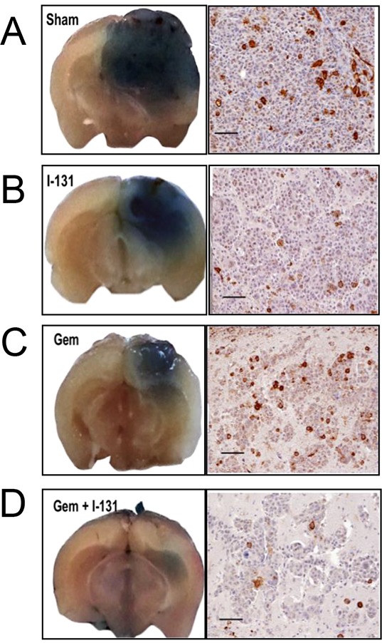
Treating breast cancer brain metastases (BCBMs) is challenging. Na+/I- symporter (NIS) expression in BCBMs would permit their selective targeting with radioiodide (131I-). We show impressive enhancement of tumor response by combining131I- with gemcitabine (GEM), a cytotoxic radiosensitizer. Nude mice mammary fat-pad (MFP) tumors and BCBMs were generated with braintropic MDA-MB-231Br cells transduced with bicistronically-linked NIS and firefly luciferase cDNAs. Response was monitored in vivo via bioluminescent imaging and NIS tumor expression.131I-/GEM therapy inhibited MFP tumor growth more effectively than either agent alone. BCBMs were treated with: high or low-dose GEM (58 or 14.5 mg/Kg×4); 131I- (1mCi or 2×0.5 mCi 7 days apart); and 131I-/GEM therapy. By post-injection day (PID) 25, 82-86% of controls and 78-83% of 131I--treated BCBM grew, whereas 17% low-dose and 36% high-dose GEM regressed. The latter tumors were smaller than the controls with comparable NIS expression (~20% of cells). High and low-dose 131I-/ GEM combinations caused 89% and 57% tumor regression, respectively. High-dose GEM/131I- delayed tumor growth: tumors increased 5-fold in size by PID45 (controls by PID18). Although fewer than 25% of cells expressed NIS, GEM/131I- caused dramatic tumor regression in NIS-transduced BCBMs. This effect was synergistic, and supports the hypothesis that GEM radiosensitizes cells to 131I-.

Keywords: breast cancer brain metastases (BCBMs); radioiodide therapy; sodium/iodide symporter (NIS).


Conflict of interest statement

CONFLICTS OF INTEREST

None of the authors have any conflicts of interest to disclose with respect to the research described herein.

Figures

Figure 1
Figure 1. Cell characterization and cytotoxicity studies
MDA-MB-231Br cells were stably transduced with a lentiviral vector containing human NIS cDNA linked via a bicistronic construct to the firefly luciferase 2 cDNA (NIS-IRES-luc2, NIL), driven by a CMV promoter. A. Immunoblot analysis of membrane fractions (15 μg) derived from transduced control MDCK cells, MDA-MB-231Br-NIL (lanes 1 and 2) and non-transduced MDA-MB-231Br (lane 3). Distinct bands corresponding to NIS protein are seen in lanes 1 and 2, but completely absent in 3. B. Functional expression of NIS was measured by quantitating 99mTcO4 uptake in single-cell-derived clonal populations. C. Functional expression of luciferase was measured using in vitro bioluminescence. Cell number correlates linearly with in vitro bioluminescence (r2 = 0.995). D–E. In vitro cytotoxicity in MDA-MB-231Br-NIL cells exposed to increasing doses of 131I (0, 40, 200, or 400 μCi) for 7 hours. [D] Representative photographs of a131I clonogenic assay and [E] surviving fraction or colony-forming efficiency determined 10 to 14 days later (mean ± SD; * p < 0.05; **** p < 0.0001 post hoc Tukey's multiple comparisons test). F. Dose-dependent effect of gemcitabine (GEM) on cell survival. Cell viability determined by MTS assay after a 48-hour incubation of 0.0001 to 1000μM GEM relative to HBSS-treated control. The concentration of the drug yielding half-maximal response (EC50) was estimated to be 1.44 ± 0.76 μM for the transduced cells and 0.588 ± 0.065 μM for the parental cells.
Figure 2
Figure 2. Radioiodide uptake in MDA-MB-231Br-NIL MFP and BCBM xenografts
A. Tissue biodistribution of 40 μCi 123I i.v. at 2, 4, and 6 hours in mice bearing NIS-expressing MDA-MB-231Br-NIL MFP xenografts. Data represent the mean ± SD of 123I uptake expressed as the percentage of the injected dose per tissue or organ (% ID/g). MFP tumors show peak accumulation of 16.8 ± 8.2 %ID/g at 2 hours decreasing to about half at 6 hours and exceeding the stomach at the earlier timepoints. B. Radioiodide uptake in brain 1 hour post-injection of 123I demonstrates that brains with tumors take up 13.6 times more 123I than normal brain tissue (n = 3). C. Immunoblot of NIS expression in MFP and BCBM xenografts shows a predominant band corresponding to the mature polypeptide and a band that migrates faster, corresponding to the partially glycosylated polypeptide. Immunohistochemical analysis of NIS expression in MFP and BCBM xenografts on day 11 post-implantation. D. Immunoblot of NIS expression in human primary triple negative breast cancer [TNBC] (*BC), and of a protein lysate (2 μg) obtained from MDCK transfected with hNIS used as a positive control showing the same electrophoretical pattern. This same tumor was minced in RPMI tissue culture and implanted into the MFP in a NOD SCID mouse to generate a patient-derived orthotopic xenograft (PDOX). Comparable NIS immunoreactivity is observed in the human TNBC and corresponding PDOX. E. NIS expression was also assessed by IHC on tissue sections of a TNBC BCBM patient is shown.
Figure 3
Figure 3. Effects of gemcitabine, 131I, and combination treatment on MDA-MB-231Br –NIL MFP tumors
MFP xenograft tumors grew over the course of a 50-day period without treatment (Saline, blue solid line), whereas growth was inhibited in tumors treated with GEM (58 mg/Kg x4, red line) or 1 mCi 131I (black line). Dual therapy, GEM 58 mg /kg × 4) plus 1 mCi 131I, induced greater tumor regression (green line). Each data point represents the average of results from at least five mice and the corresponding SEM.
Figure 4
Figure 4. Effects of gemcitabine, 131I, and combination treatment on MDA-MB-231Br –NIL BCBM
A–D. Mice were injected into the right cerebral cortex with 0.2 × 10⁶ cells. Tumor BLI was monitored bi-weekly starting on day 7. Animals with tumors showing exponential growth by day 10 (baseline) were randomly assigned to different treatment groups: [A] saline solution alone (dotted arrows); [B] 131I 1mCi (black arrow) on day 11 and saline other days; [C] GEM 58 mg/Kg (gray arrows) on days 10, 13, 16, 19; [D] dual therapy: GEM on days 10,13,16,19 and 131I (black arrow) on day 11. Pseudo-color BLI images show a representative animal from each group.
Figure 5
Figure 5. Effectiveness of the dual treatment
Representative photographs of whole-brain coronal sections from mice infused with Evans blue prior to sacrifice, highlighting the location of metastases (left). Staining ranged from intensely blue in the larger tumors from sham or saline-treated mice A. to smaller 131I- and GEM-treated tumors B. and C. respectively. Dual therapy resulted in the smallest tumors of all D. Accompanying panels show histologic sections stained with anti-NIS antibody (scale bars 100 μm). Saline-treated tumors show NIS membrane staining in ~15% of cells, whereas those treated with 131I at least doubled in size, using the BLI criterion, and <5% of their cells were NIS-positive [B]. In GEM-treated tumors that regressed [C], the proportion of NIS-positive cells was similar to that in the control group. In the GEM/131I group [D], tumors were smallest or undetectable; in the tumor shown, which was classified as stable, <5% of the cells were NIS-positive.
Figure 6
Figure 6. Effects of different treatments on tumor growth
A–B. Tumor response for each animal was classified as growth if BLI was at least twice as intense as the baseline, regression if it was half or less as intense, and stable disease if BLI remained within the 2-fold parameter on PID25. The percentage of mice in each category is shown in the bar graph. [A] Effects of the low-dose regimens [14.5 mg GEM /kg (days 10, 13, 16, 19) and 2 × 0.5 mCi 131I (days 11 and 18)], with each agent alone or both combined. [B] Effects of the high-dose regimens [GEM 58 mg /kg (days 10, 13, 16, 19) and 1 mCi 131I (day 11)], with each agent alone or both combined. C–D. Comparison of antitumor activity of low and high dose GEM, 131I, and combination treatment on MDA-MB-23-Br-NIL brain tumors at PID25. Treatments and control groups were compared by one way-analysis of variance followed by post hoc comparisons using the Dunnett's multiple-comparisons test. Statistics were performed on log-transformed data. The threshold for significance was set at P<0.05. ns P>0.05, * P≤0.05, ** P≤0.01, *** P≤0.001, **** P≤0.0001.
Figure 7
Figure 7. Dual therapy delays tumor progression
Kaplan-Meier survival analysis is shown using median survival time for each treatment group or the time required to achieve 5 times the bioluminescence compared to its respective baseline on PID10. Sham or saline-treated [gray], 131I [blue], GEM [green], GEM + 131I [red]. The threshold for significance was set at p < 0.05.

Similar articles

Cited by

References

    1. Dawood S, Gonzalez-Angulo AM. Progress in the biological understanding and management of breast cancer-associated central nervous system metastases. Oncologist AlphaMed Press. 2013;18:675–84. - PMC - PubMed
    1. Lin NU, Amiri-Kordestani L, Palmieri D, Liewehr DJ, Steeg PS. CNS metastases in breast cancer: old challenge, new frontiers. Clin Cancer Res. American Association for Cancer Research. 2013;19:6404–18. - PMC - PubMed
    1. Muldoon LL, Soussain C, Jahnke K, Johanson C, Siegal T, Smith QR, Hall WA, Hynynen K, Senter PD, Peereboom DM, Neuwelt EA. Chemotherapy delivery issues in central nervous system malignancy: a reality check. J Clin Oncol. 2007;25:2295–305. - PubMed
    1. Wapnir IL, van de Rijn M, Nowels K, Amenta PS, Walton K, Montgomery K, Greco RS, Dohan O, Carrasco N. Immunohistochemical profile of the sodium/iodide symporter in thyroid, breast, and other carcinomas using high density tissue microarrays and conventional sections. J Clin Endocrinol Metab. 2003;88:1880–8. - PubMed
    1. Portulano C, Paroder-Belenitsky M, Carrasco N. The Na+/I- symporter (NIS): mechanism and medical impact. Endocr Rev. 2014;35:106–49. - PMC - PubMed

MeSH terms