. 2016 Aug 2;7(31):49259-49267.
doi: 10.18632/oncotarget.10272.

Concerted action of IFN-α and IFN-λ induces local NK cell immunity and halts cancer growth

Affiliations

Concerted action of IFN-α and IFN-λ induces local NK cell immunity and halts cancer growth

Ahmed Lasfar et al. Oncotarget. .

Abstract

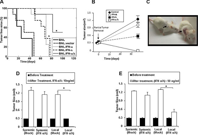
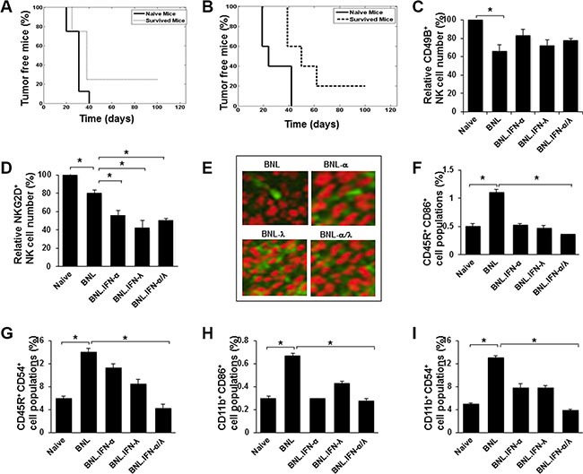
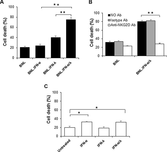
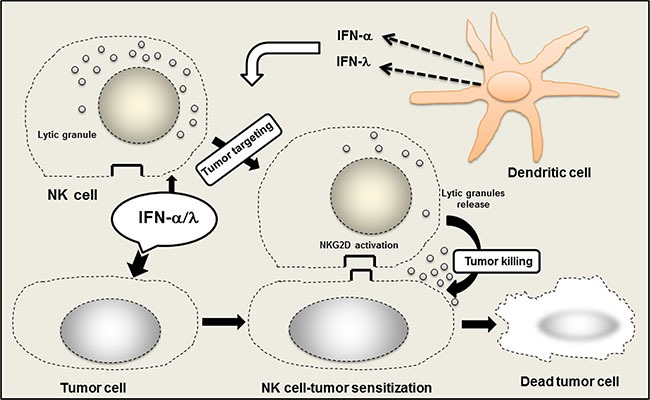
Hepatocellular carcinoma (HCC) is the most prevalent type of liver cancer. No significant improvement has been reported with currently available systemic therapies. IFN-α has been tested in both clinic and animal models and only moderate benefits have been observed. In animal models, similar modest antitumor efficacy has also been reported for IFN-λ, a new type of IFN that acts through its own receptor complex. In the present study, the antitumor efficacy of the combination of IFN-α and IFN-λ was tested in the BNL mouse hepatoma model. This study was accomplished by using either engineered tumor cells (IFN-α/IFN-λ gene therapy) or by directly injecting tumor-bearing mice with IFN-α/IFN-λ. Both approaches demonstrated that IFN-α/IFN-λ combination therapy was more efficacious than IFN monotherapy based on either IFN-α or IFN-λ. In complement to tumor surgery, IFN-α/IFN-λ combination induced complete tumor remission. Highest antitumor efficacy has been obtained following local administration of IFN-α/IFN-λ combination at the tumor site that was associated with strong NK cells tumor infiltration. This supports the use of IFN-α/IFN-λ combination as a new cancer immunotherapy for stimulating antitumor response after cancer surgery.

Keywords: HCV; IFN therapy; IFN-α/IFN-λ combination; hepatocellular carcinoma; tumor immunity.


Conflict of interest statement

The authors disclose no potential conflicts of interest.

Figures

Figure 1
Figure 1. Synergisitic effects of IFN-α and IFN-λ on in vivo BNL tumor growth
(A) Syngeneic BALB/c mice (n = 8) were injected s.c. in the flank with 106 BNL, BNL.vector, BNL.IFN-λ, BNL.IFN-α cells, or a 50:50 combination of both IFN-producing cells. Data in the Kaplan-Meier survival curves are shown as percentage of tumor free mice. BNL.IFN-α or BNL.IFN-λ versus BNL.IFN-α/λ cells (*) p < 0.05 (one-way ANOVA). (B) Mice (n = 5 per group) were inoculated with 106 parental BNL cells at the back. After tumor growth (1 cm3), partial tumor removal was performed (0.3 cm3 left). Mice were then treated with 10 ng of either IFN-α or IFN-λ alone, or a combination of IFN-α and IFN-λ (50% of each dose) at the tumor surgery site, starting two days post-surgery, three days a week for two weeks; and tumor growth was monitored. Data are presented as the mean tumor volume ± SE (n = 5). (C) Representative example showing a mouse, treated with IFN-α and IFN-λ combination (Left) and control (Right). Arrows indicate the tumor site and area of treatment with IFN-α and IFN-λ combination or control (Mock). (D and E) Mice (n = 5 per group) were inoculated with 106 parental BNL cells at the neck. When the tumor reached around 1 cm3, partial tumor removal was performed. Mice were then treated locally at the tumor inoculation site at the neck or systemically (i. p) with either 10 ng (d) or 50 ng (e) of a combination of IFN-α and IFN-λ (50% of each IFN dose), three days a week for two weeks and starting two days post-surgery. Experiments are repeated 3 times and data presented as the mean tumor volume ± SE. (*) p < 0.05. p value determined by Mann-Whitney U test.
Figure 2
Figure 2. Antitumor immunity induced by the IFN-α/λ combination
(A and B) Survived mice in the experiments represented in Figure 1A (n = 4) and Figure 1C (n = 5), respectively, were re-challenged with a s.c. injection at the original tumor inoculation site (left flank) with 106 parental BNL cells. Mice were followed for 3 months and percentage of tumor free mice was determined. (C and D.) Blood was isolated from naive mice or mice at 15 days post injection with 106 of parental BNL or engineered BNL cells, BNL.IFN-α, BNL.IFN-λ and the combination of BNL.IFN-α and BNL.IFN-λ (BNL.IFN-α/λ) cells (50% of each). The number of circulatory CD49B+ NK cells (c), CD49B+/NKG2D+ NK cells (d) was determined by flow cytometry in each group of mice. Relative cell numbers (%) are shown as the mean ± SE (n = 3 mice per data point). Relative cell numbers (%) are shown as the mean ± SE (n = 3 tumors per data point). (E) Immunohistochemical staining with green fluorescence of NK cells (CD49B+), infiltrating parental or engineered BNL hepatoma tumors. Images were taken on confocal fluorescence microscope (x250). (FI) Assessment of circulatory CD45R+/CD86+ (f), CD45R+/CD54+ (g), CD11b+/CD86+ (h) and CD11b+/CD54+ (i) populations in mice blood. Blood was isolated from naive mice or mice at 15 days post injection with 106 of parental BNL or engineered BNL cells, BNL.IFN-α, BNL.IFN-λ and the combination of BNL.IFN-α and BNL.IFN-λ (BNL.IFN-α/λ) cells (50% of each). The number of cells was determined in each group of mice and presented as relative cell numbers (%) and shown as the mean ± SE (n = 3 mice per data point). P values are calculated using Mann-Whitney U test. (*) p < 0.05.
Figure 3
Figure 3. NK cells induced tumor immunotoxicity
(A) NK cells were evaluated for their tumor toxicity on target cells, parental BNL or engineered BNL cells, BNL.IFN-α, BNL.IFN-λ and BNL.IFN-α/λ cells. After co-culture of NK cells (purified from spleen of naïve mice) with target cells, cells were harvested, PI stained and the amount of cell death was assessed by FACS as indicated in Supplementary Figure 1. Dead cells (%) are shown as mean ± SE (n = 3 by data points). (B) Effect of the blocking anti-NKG2D antibody (C7) on the promotion of tumor toxicity using IFN-α/λ-secreting BNL cells. After one hour incubation of anti-NKG2D or isotype control antibody with NK cells, tumor cytotoxicity was evaluated. (C) Effect of pretreatment of NK cells with IFN on the induction of NK cells cytotoxicity on parental BNL tumors. Dead cells (%) are shown as mean ± SE (n = 3 by data points, (**) p < 0.01, (*) p < 0.05). P values are calculated using Mann-Whitney U test.
Figure 4
Figure 4. Role of NK cells in the promotion of anticancer efficacy of IFN-α/λ combination
Within the tumor site, IFN-α/λ induced NK cells activation, mostly via tumor cell sensitization to NK cells. Concerted action of IFN-α and IFN-λ promotes NK cell tumor targeting and induces tumor eradication. IFN-α/λ is released in the tumor site by direct injection or through IFN secretion by dendritic cells (DCs) either conventional or plasmacytoid DCs. Activation of NKG2D system by IFN-α/λ appears crucial within the tumor microenvironment for the promotion of local NK cell tumor surveillance.

Similar articles

Cited by

References

    1. Sherman M. Hepatocellular carcinoma: epidemiology, surveillance, and diagnosis. Seminars in liver disease. 2010;30:3–16. - PubMed
    1. Georgiades C, Geschwind JF, Harrison N, Hines-Peralta A, Liapi E, Hong K, Wu Z, Kamel I, Frangakis C. Lack of Response after Initial Chemoembolization for Hepatocellular Carcinoma: Does It Predict Failure of Subsequent Treatment? Radiology. 2012 - PMC - PubMed
    1. Melloul E, Lesurtel M, Carr BI, Clavien PA. Developments in liver transplantation for hepatocellular carcinoma. Seminars in oncology. 2012;39:510–521. - PubMed
    1. Shimomura S, Ikeda N, Saito M, Ishii A, Takashima T, Sakai Y, Yoshikawa S, Aizawa N, Tanaka H, Iwata Y, Enomoto H, Imanishi H, Yamamoto T, et al. Long-term interferon therapy after radiofrequency ablation is effective in treating patients with HCV-associated hepatocellular carcinoma. Hepatol Int. 2010;5:559–566. - PMC - PubMed
    1. Lin SM, Yu ML, Lee CM, Chien RN, Sheen IS, Chu CM, Liaw YF. Interferon therapy in HBeAg positive chronic hepatitis reduces progression to cirrhosis and hepatocellular carcinoma. J Hepatol. 2007;46:45–52. - PubMed