. 2016 Jul 1:6:27583.
doi: 10.1038/srep27583.

Abrogating ClC-3 Inhibits LPS-induced Inflammation via Blocking the TLR4/NF-κB Pathway

Affiliations

Abrogating ClC-3 Inhibits LPS-induced Inflammation via Blocking the TLR4/NF-κB Pathway

Nan-Lin Xiang et al. Sci Rep. .

Abstract

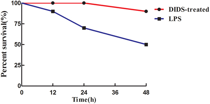
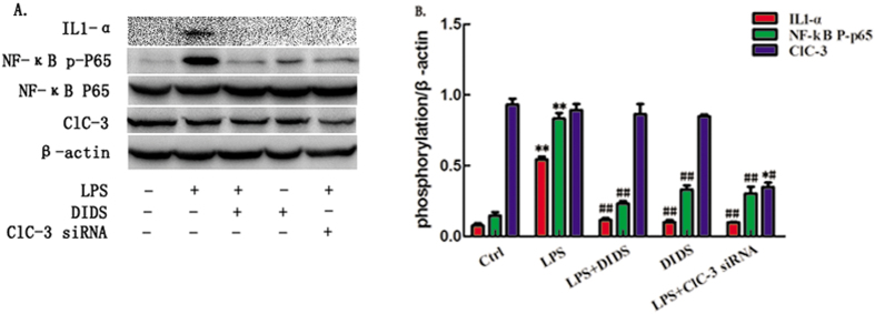
This study investigated the function of a chloride channel blocker, DIDS. Both in vitro and in vivo studies found that DIDS significantly inhibits lipopolysaccharide (LPS)-induced release of proin flammatory cytokines. Here, we show that DIDS inhibits LPS-induced inflammation, as shown by downregulation of inflammatory cytokines via inhibition of the TLR4/NF-κB pathway. Furthermore, we show that ClC-3siRNA transfection reduces LPS-induced pro-inflammation in Raw264.7 cells, indicating that ClC-3 is involved in the inhibitory effect of DIDS during LPS-induced cytokines release. In vivo, DIDS reduced LPS-induced mortality, decreased LPS-induced organic damage, and down-regulated LPS-induced expression of inflammatory cytokines. In sum, we demonstrate that ClC-3 is a pro-inflammatory factor and that inhibition of ClC-3 inhibits inflammatory induction both in vitro and in vivo, suggesting that ClC-3 is a potential anti-inflammatory target.


Figures

Figure 1
Figure 1. Chloride Channel Blocker DIDS Down-regulates LPS-induced Production of Inflammatory Cytokines in RAW264.7 Cells.
(A) Inflammatory cytokine array analysis of IL1-α, IL1-β, IL-6 and TNF-α. The expression of these inflammatory cytokines is highly induced by LPS, but down-regulated by DIDS and ClC-3 siRNA. (B–I) Western -blotting of IL1-α, IL1-β, IL-6 and TNF-α in RAW264.7 cells in each group. Quantification of western -blotting band intensity of IL1-α, IL1-β, IL-6 and TNF-α corrected for the intensity of β-actin with image-analysis software. (**P < 0.01 vs control, #P < 0.05, ##P < 0.01 vs LPS; n = 3).
Figure 2
Figure 2. ClC-3siRNA Transfection Reduces LPS-induced Production of Cytokine IL1-α.
(A) Cell lysates of ClC-3 siRNA and DIDS in each group were prepared and immunoblotted with ClC-3 and IL1-α antibodies. (B) Quantification of western-blotting band intensity fpr IL1-α corrected for the intensity of β-actin with image-analysis software. (*P < 0.05 vs control, #P < 0.05 vs LPS; n = 4).
Figure 3
Figure 3. Chloride Channel Blocker DIDS and ClC-3 siRNA Transfection Reduces LPS-Induced Expression of TLR4 in Macrophages.
(A) Cell lysates of ClC-3 siRNA and DIDS in each group were prepared and immunoblotted with ClC-3, TLR4 and IL1-α antibodies. (B) Quantification of western -blotting band intensity of IL1-α and TLR4, respectively, corrected for the intensity of β-actin with image-analysis software. (*P < 0.05, **P < 0.01 vs control, #P < 0.05, ##P < 0.01 vs LPS; n = 3).
Figure 4
Figure 4. Chloride Channel Blocker DIDS and ClC-3 siRNA transfection Inhibit LPS-induced Production of Inflammatory Cytokines via NF-κB Signaling Pathway.
(A) RAW264.7 cells were pretreated with either DIDS or ClC-3siRNA, followed by stimulation with LPS for the indicated time points. NF-κB phosphorylation was determined by Western -blotting. (B) Quantification of western -blotting band intensity of IL1-α and p-p65, corrected for the intensity of β-actin with image-analysis software. (**P < 0.01 vs control, #P < 0.05, ##P < 0.01 vs LPS; n = 3).
Figure 5
Figure 5. Chloride Channel Blocker DIDS Reduces LPS-Induced Mortality in Mice.
DIDS was injected i.p. 30 min before coinjection with LPS. The survival rate was measured after 12 h, 24 h and 48 h.
Figure 6
Figure 6. Chloride Channel Blocker DIDS Reduces LPS-induced Organ Damage in Mice.
(A,B) DIDS was i.p.-injected into mice 6 h before LPS injection. Serum samples were collected 6 h after coinjection, and ALT and BUN activity in the serum were determined. (C,D) H&E staining of hepatorenal tissue from each group of mice at different magnifications. Livers of LPS-treated mice (Fig. 5C). As shown by the black arrow, the topical area of the liver tissue shows many fat particles. As shown by the red arrow, the hepatic veins are filled with red blood cells. However, in the livers of DIDS/LPS-treated mice, a small amount of hepatic sinus is slightly dilated and liver damage is decreased. The same expression is observed in the kidney tissue, of LPS-infused mice (Fig. 5D). As shown by the black arrow, there is a decrease in the number of glomerular capillary loop, and glomerular atrophy. As shown by the green arrow, there is topical proliferation of glomerular epithelial cells. As shown by the blue arrow, the nucleus of renal tubular epithelial cells are condensed, undergoing necrocytosis, and there is degradation of the cytoplasm protein and the structure of the kidney tubules has disappeared. As shown by the red arrow, there is dilatation and hyperemia of the blood capillary in the renal tubulointerstitial space. In the kidney tissue of DIDS/LPS-treated mice, the structure of the glomerulus and kidney tubules are integrated, with only an small amount of topical proliferation in glomerular epithelial cells, as the green arrow shown. (**P < 0.01 vs control, #P < 0.05, ##P < 0.01 vs LPS; n = 5).
Figure 7
Figure 7. Chloride Channel Blocker DIDS Down-regulates LPS-induced Expression of Inflammatory Cytokines in Mice.
DIDS was injected i.p. 30 min before coinjection with LPS. (A) Serum samples in each group were collected at 6 h and IL1-α was measured by ELISA. (B) IL1-β was measured by ELISA. (C) IL-6 was measured by ELISA. (D) TNF-α was measured by ELISA. (*P < 0.05, **P < 0.01 vs control, #P < 0.05 vs LPS; n = 3).
Figure 8
Figure 8. Chloride Channel Blocker DIDS Down-regulates LPS-Induced Production of IL1-α and NF-κB Phosphorylated P65 in Primary Macrophages.
(A) Western -blotting of IL1-α and NF-κB phosphorylated P65 in primary macrophages. (B) Quantification of western-blotting band intensity of IL-1α and p-p65, corrected for the intensity of β-actin with image-analysis software. (**P < 0.01 vs control, #P < 0.05, ##<0.01 vs LPS; n = 3).

References

    1. Lei Y. L., Wang K. & Deng L. F. Redox regulation of inflammation: old elements, a new story. Med Res Rev. 35, 306–340 (2015). - PubMed
    1. Bhatia M. et al. H2S and Inflammation: An Overview. Handb Exp Pharmacol. 230, 165–180 (2015). - PubMed
    1. Davies L. C. & Taylor P. R. Tissue-resident macrophages: then and now. Immunology 144, 541–548 (2015). - PMC - PubMed
    1. Wyns H. et al. In vivo porcine lipopolysaccharide inflammation models to study immunomodulation of drugs. Vet Immunol Immunopathol. 166, 58–69 (2015). - PubMed
    1. Liu A. H. et al. DIDS attenuates staurosporine-induced cardiomyocyte apoptosis by PI3K/Akt signaling pathway: activation of eNOS/NO and inhibition of Bax translocation. Cell Physiol Biochem. 22, 177–186 (2008). - PubMed

Publication types

MeSH terms