Review
. 2016 Sep;22(5):535-60.
doi: 10.1093/humupd/dmw022. Epub 2016 Jun 30.

Novel concepts on pregnancy clocks and alarms: redundancy and synergy in human parturition

Affiliations
Review

Novel concepts on pregnancy clocks and alarms: redundancy and synergy in human parturition

Ramkumar Menon et al. Hum Reprod Update. 2016 Sep.

Abstract

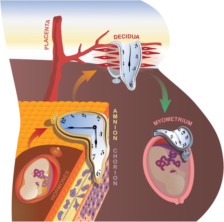
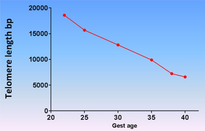
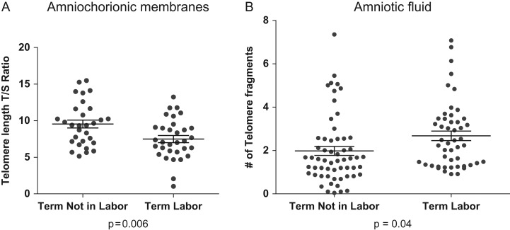
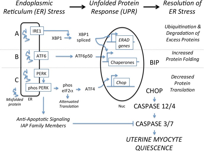
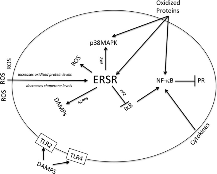
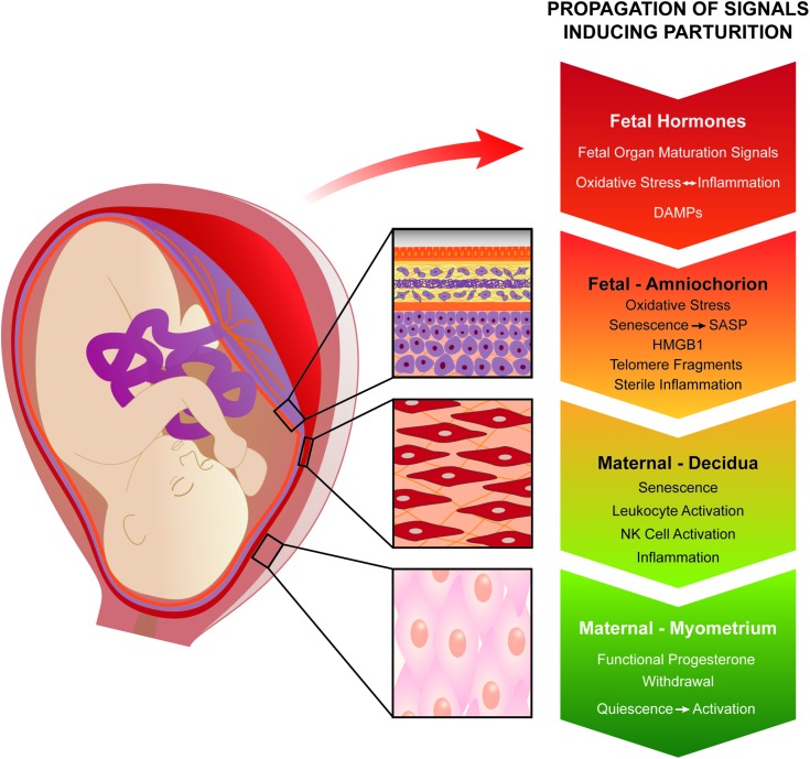
The signals and mechanisms that synchronize the timing of human parturition remain a mystery and a better understanding of these processes is essential to avert adverse pregnancy outcomes. Although our insights into human labor initiation have been informed by studies in animal models, the timing of parturition relative to fetal maturation varies among viviparous species, indicative of phylogenetically different clocks and alarms; but what is clear is that important common pathways must converge to control the birth process. For example, in all species, parturition involves the transition of the myometrium from a relaxed to a highly excitable state, where the muscle rhythmically and forcefully contracts, softening the cervical extracellular matrix to allow distensibility and dilatation and thus a shearing of the fetal membranes to facilitate their rupture. We review a number of theories promulgated to explain how a variety of different timing mechanisms, including fetal membrane cell senescence, circadian endocrine clocks, and inflammatory and mechanical factors, are coordinated as initiators and effectors of parturition. Many of these factors have been independently described with a focus on specific tissue compartments.In this review, we put forth the core hypothesis that fetal membrane (amnion and chorion) senescence is the initiator of a coordinated, redundant signal cascade leading to parturition. Whether modified by oxidative stress or other factors, this process constitutes a counting device, i.e. a clock, that measures maturation of the fetal organ systems and the production of hormones and other soluble mediators (including alarmins) and that promotes inflammation and orchestrates an immune cascade to propagate signals across different uterine compartments. This mechanism in turn sensitizes decidual responsiveness and eventually promotes functional progesterone withdrawal in the myometrium, leading to increased myometrial cell contraction and the triggering of parturition. Linkage of these processes allows convergence and integration of the gestational clocks and alarms, prompting a timely and safe birth. In summary, we provide a comprehensive synthesis of the mediators that contribute to the timing of human labor. Integrating these concepts will provide a better understanding of human parturition and ultimately improve pregnancy outcomes.

Keywords: NK cells; aging; corticosteroids; endoplasmic reticulum; labor and delivery; oxidative stress; p38MAPK; progesterone receptors; senescence; unfolded proteins.


Figures

Figure 1
Figure 1
Proposed system of multiple biological clocks and alarms to optimize the duration of pregnancy. During pregnancy, at least four such timing mechanisms exist and these can operate in parallel or in series. Neuroendocrine fetal hormones and placental factors, particularly those modulating cortisol productions, have been classically invoked and are reviewed under ‘The endocrine clock’. The novel concept of natural fetal membrane cell senescence is developed under ‘Fetal membrane clock: a source of parturition signals’, where several testable, amnion-derived sterile inflammatory mediators are proposed. The decidual clock serves at the critical vascular feto–maternal interface (The decidual clock: inflammation and senescence). Ultimately, the myometrium becomes progesterone resistant and is activated to generate coordinated, forceful contractions, leading to fetal expulsion (Myometrial clock: progesterone and inflammation). Oxidative, nitrosative, folded-protein and ER stress pathways (Myometrial stretch inputs and activation of parturition) also contribute to the molecular regulation of labor onset. Finally, redundancy and synergy provide assurance against prolonged pregnancy duration, whereas premature activation of one or more pathways can trigger early delivery. Understanding the integrated actions of these systems will inform new clinical approaches to the control of labor onset.
Figure 2
Figure 2
Fetal telomere shortening during pregnancy. Fetal leukocyte DNA telomere analysis determined progressive reduction in telomere length as gestation progresses. The longest telomere length was seen at 22 weeks and the shortest at term. This is indicative of a telomere-dependent senescence process in the fetal compartment.
Figure 3
Figure 3
Telomere length changes in fetal membranes and telomere fragments in amniotic fluid at term. Telomere length reduction in fetal membranes in term laboring membranes compared with term not-in-labor membranes. (A) At term, the transition from not-in-labor to labor is associated with telomere length reduction in fetal membranes. (B) The decrease in fetal membrane telomere length is associated with an increase in telomere fragments in the amniotic fluid of women undergoing labor and delivery.
Figure 4
Figure 4
The ERSR-UPR signaling cascade. IRE1, serine/threonine-protein kinase/endoribonuclease inositol-requiring enzyme 1; ATF6, activating transcription factor 6; PERK, protein kinase RNA-like endoplasmic reticulum kinase; ERSR, endoplasmic reticulum stress response; eIF2α, eukaryotic initiation factor 2 alpha; XBP1, X-box binding protein 1; BiP, binding immunoglobulin protein; CHOP, CCAAT-enhancer-binding protein homologous protein. This figure is adapted from in vivo cellular adaptation to ER stress: survival strategies with double-edged consequences (Tsang et al., 2010).
Figure 5
Figure 5
Modulation of the uterine ERSR-UPR by senescence-associated secreted factors. ROS, reactive oxygen species; PR, progesterone receptor; DAMPs, damage associated molecular patterns.
Figure 6
Figure 6
Propagation of fetal signals that will initiate parturition. In this review, we project fetal membrane senescence as one of the major contributors of initiation of human parturition. Natural and physiologic aging of fetal membranes mediated by telomere-dependent senescence and senescence-associated inflammation (contributed by senescence associated secretory phenotype [SASP] and damage associated molecular patterns [DAMP]) increases the overall inflammatory load in fetal membranes. These inflammatory signals get propagated to decidual layer, causing leukocyte and natural killer (NK) cell activation, causing inflammatory overload in the decidua. Fetal membrane and decidual inflammatory overload transition the quiescent myometrium to an active state of functional progesterone withdrawal.

References

    1. Aagaard K, Ma J, Antony KM, Ganu R, Petrosino J, Versalovic J. The placenta harbors a unique microbiome. Sci Transl Med 2014;6:237ra65. - PMC - PubMed
    1. Abdel-Hafiz HA, Horwitz KB. Post-translational modifications of the progesterone receptors. J Steroid Biochem Mol Biol 2014;140:80–89. - PMC - PubMed
    1. Abdel-Hafiz H, Takimoto GS, Tung L, Horwitz KB. The inhibitory function in human progesterone receptor N termini binds SUMO-1 protein to regulate autoinhibition and transrepression. J Biol Chem 2002;277:33950–33956. - PubMed
    1. ACOG Committee Opinion No 579: Definition of term pregnancy. Obstet Gynecol 2013;122:139–1140. - PubMed
    1. Adams Waldorf KM, Singh N, Mohan AR, Young RC, Ngo L, Das A, Tsai J, Bansal A, Paolella L, Herbert BR et al. . Uterine overdistention induces preterm labor mediated by inflammation: observations in pregnant women and nonhuman primates. Am J Obstet Gynecol 2015;213:830.E1-830.19. - PMC - PubMed