. 2016 Jul 31;428(15):3043-57.
doi: 10.1016/j.jmb.2016.06.014. Epub 2016 Jun 27.

Noncanonical Myo9b-RhoGAP Accelerates RhoA GTP Hydrolysis by a Dual-Arginine-Finger Mechanism

Affiliations

Noncanonical Myo9b-RhoGAP Accelerates RhoA GTP Hydrolysis by a Dual-Arginine-Finger Mechanism

Fengshuang Yi et al. J Mol Biol. .

Abstract

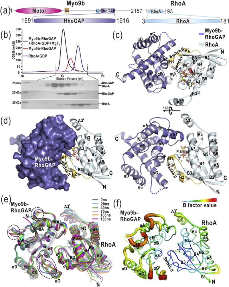
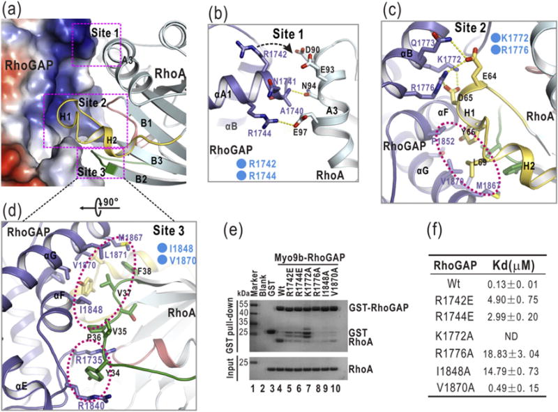
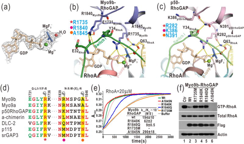
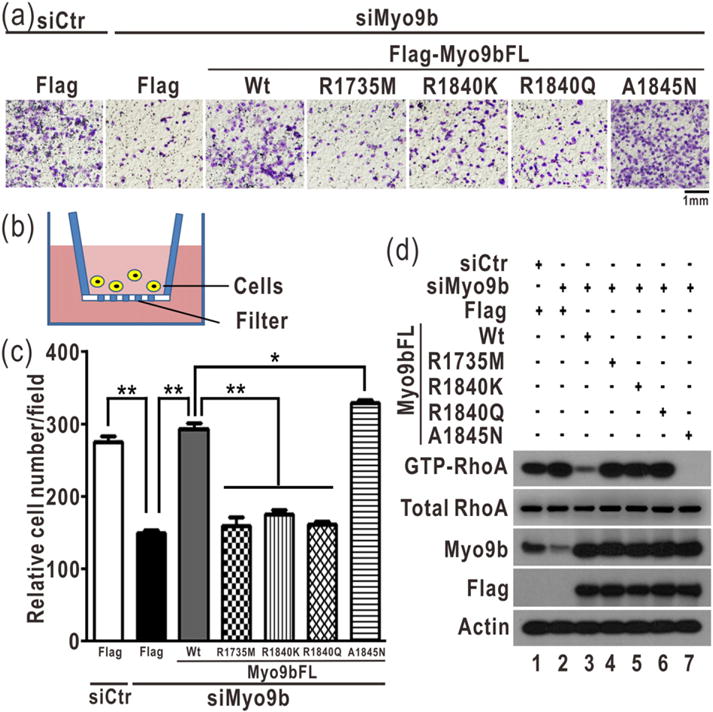
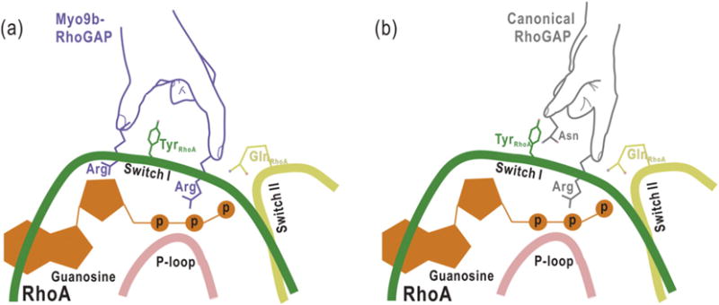
The GTP hydrolysis activities of Rho GTPases are stimulated by GTPase-activating proteins (GAPs), which contain a RhoGAP domain equipped with a characteristic arginine finger and an auxiliary asparagine for catalysis. However, the auxiliary asparagine is missing in the RhoGAP domain of Myo9b (Myo9b-RhoGAP), a unique motorized RhoGAP that specifically targets RhoA for controlling cell motility. Here, we determined the structure of Myo9b-RhoGAP in complex with GDP-bound RhoA and magnesium fluoride. Unexpectedly, Myo9b-RhoGAP contains two arginine fingers at its catalytic site. The first arginine finger resembles the one within the canonical RhoGAP domains and inserts into the nucleotide-binding pocket of RhoA, whereas the second arginine finger anchors the Switch I loop of RhoA and interacts with the nucleotide, stabilizing the transition state of GTP hydrolysis and compensating for the lack of the asparagine. Mutating either of the two arginine fingers impaired the catalytic activity of Myo9b-RhoGAP and affected the Myo9b-mediated cell migration. Our data indicate that Myo9b-RhoGAP accelerates RhoA GTP hydrolysis by a previously unknown dual-arginine-finger mechanism, which may be shared by other noncanonical RhoGAP domains lacking the auxiliary asparagine.

Keywords: GTPase-activating proteins; Myo9b; Rho GTPases; RhoA; RhoGAP domain.


Figures

Fig. 1
Fig. 1
The overall structure of the Myo9b-RhoGAP/RhoA complex. (a) Domain organization of Myo9b and RhoA. Myo9b contains an N-terminal motor domain followed by four IQ motifs and a C1, and a RhoGAP domain at the C terminus. RhoA contains a Rho GTPase domain. (b) Analytical gel-filtration analysis of the formation of the Myo9b-RhoGAP/RhoA complex in the presence of MgF3. (c) A ribbon diagram of the structure of the Myo9b-RhoGAP/RhoA complex. Myo9b-RhoGAP and RhoA are colored in blue and cyan, respectively. The secondary structures of Myo9b-RhoGAP and RhoA are labeled and both N and C termini are also marked. The Switch I and II and P-loop of RhoA, colored in green, yellow, and pink, respectively, construct the nucleotide-binding pocket in which GDP, MgF3, and Mg2+ are located. (d) A combined surface and ribbon representation of the Myo9b-RhoGAP/RhoA complex structure. Myo9b-RhoGAP is in the surface representation (colored in blue), and RhoA is in the ribbon representation (colored in cyan). The figure shows that RhoA is cradled in the expanded concave target-binding groove of Myo9b-RhoGAP. (e) Superposition of the snapshots of a representative simulation of the Myo9b-RhoGAP/RhoA complex with simulation time indicated. The flexible loops of Myo9b-RhoGAP that exhibit dynamic conformations during the simulations are highlighted by dashed circles. (f) A diagrammatic representation of the complex structure with variable radii according to different B-factors. Consistent with data from the MD simulations, the flexible loops of Myo9b-RhoGAP have higher B-factors.
Fig. 2
Fig. 2
The interaction interface between Myo9b-RhoGAP and RhoA. (a) A combined surface-and-ribbon representation showing the interaction interface between Myo9b-RhoGAP and RhoA. RhoA is presented in the ribbon representation (colored in cyan), and Myo9b-RhoGAP is shown in the surface representation. In this surface drawing, the positive and negative charge potentials are colored in blue and red, respectively. Notably, the target-binding groove of Myo9b-RhoGAP is predominantly positively charged. The three interaction sites are also highlighted by dashed boxes. (b–d) A combined ribbon-and-stick model illustrates the three interaction sites (Site 1 to 3) between Myo9b-RhoGAP and RhoA. In this drawing, Myo9b-RhoGAP and RhoA are colored following the color patterns of Fig. 1c, and the side chains of the residues involved in the interaction are shown as sticks. Hydrogen bonds and salt bridges are indicated by dashed lines. Hydrophobic packing clusters are also highlighted by dashed circles. (e) GST pull-down analysis of the interactions between the Myo9b-RhoGAP mutants and RhoA. The GST protein alone was used as the negative control. As compared to the wild-type protein, all the mutations in Myo9b-RhoGAP severely impaired the binding between Myo9b-RhoGAP and RhoA. (f) A summary of the binding affinities between Myo9b-RhoGAP (or its mutants) and RhoA. Due to the poor behavior of the K1722A mutant in solution, the binding affinity of this mutant for RhoA could not be determined.
Fig. 3
Fig. 3
Myo9b-RhoGAP contains two arginine fingers for RhoA GTP hydrolysis. (a) A close-up view of GDP and MgF3 in the complex structure by a stick model representation. The electron density map (2Fo-Fc map) of GDP, MgF3, Mg2+, and a putative nucleophilic water molecule is shown and contoured at 1.5 σ level. (b) A combined ribbon-and-stick model illustrates the catalytic site in the structure of the Myo9b-RhoGAP/RhoA complex. In this drawing, Myo9b-RhoGAP and the Switch I and II and P-loop of RhoA are colored in blue, green, yellow, and pink, respectively, and the residues essential for catalysis are shown as sticks. Hydrogen bonds and salt bridges are indicated by dashed lines. (c) A combined ribbon-and-stick model illustrates the catalytic site in the structure of the p50-RhoGAP/RhoA complex (PDB code: 1OW3). p50-RhoGAP is colored in violet. The color schemes of RhoA follow that shown in panel (b), and the residues essential for catalysis are shown as sticks. (d) Two selected key regions of the structure-based sequence alignment of the RhoGAP domains from different proteins. The identical and highly conserved residues are colored in red and green, respectively. The catalytic arginine finger, the auxiliary arginine finger, and the essential asparagine in the RhoGAP domains are highlighted with a red star, purple dot, and yellow dot, respectively, at the bottom. (e) Time courses of GTP hydrolysis for RhoA (20 μM) catalyzed by Myo9b-RhoGAP and its mutants (40 nM). The Michaelis–Menten kinetic parameters (kcat/Km) are summarized as an inset. Data shown are mean values ± SD from two independent experiments. (f) Biochemical pull-down analysis of active GTP-bound RhoA with Myo9b-RhoGAP and its mutants. The active RhoA levels were measured by GST pull-down analysis with GST-RBD and analyzed by Western blotting using a specific anti-RhoA antibody. The levels of total RhoA, Flag-tagged Myo9b-RhoGAP, and the actin (as an internal loading control) in the cell lysate were also shown. Consistently, mutations of the two arginine fingers impaired the catalytic activity of Myo9b-RhoGAP, while the A1845N mutation increased that of Myo9b-RhoGAP.
Fig. 4
Fig. 4
The two arginine fingers are essential for Myo9b-mediated cell migration. (a) Transwell assay of Myo9b-mediated cell migration with Myo9b and its mutants. In this assay, the cells were stained with crystal violet and the images were taken of the underside of a transwell, as shown in panel (b). The scale bar represents 1 mm. As expected, knockdown of Myo9b severely impaired cell migration. The transfection of the wild-type Myo9b largely rescued the cell migration defect induced by knockdown, whereas the transfection of the Myo9b mutants with the mutations of the two arginine fingers did not. The A1845N mutant could rescue the cell migration defect more efficiently than the wild-type protein. (b) A schematic diagram illustrates the transwell cell migration assay. In culture media, cells could penetrate into and migrate through the filter membrane in this assay. (c) Quantification of the transwell cell migration data shown in panel (a). The relative cell number of each field was quantified for each construct. Data were presented as the mean value ± SD from three independent experiments (five different fields for each experiment), **P < 0.01, *P < 0.05. (d) Biochemical pull-down analysis of active GTP-bound RhoA with the knockdown of Myo9b and transfection of the Flag-tagged, full-length Myo9b or its mutants. All the data were presented as shown in Fig. 3f. The level of Myo9b in the cell lysate was analyzed by Western blotting using the specific anti-Myo9b antibody. Consistently, the level of active GTP-bound RhoA was increased by down-regulating Myo9b, and the level of active RhoA was decreased by transfecting the wild-type Myo9b and A1845N mutant but not the Myo9b mutants with impairment of the two arginine fingers.
Fig. 5
Fig. 5
The proposed schematic models illustrating the catalytic mechanisms for Myo9b-RhoGAP and the canonical RhoGAP domain. Myo9b-RhoGAP contains two arginine fingers at the catalytic site (a), whereas the canonical RhoGAP domain has only one arginine finger together with an auxiliary asparagine (b). Similar to the catalytic arginine finger in canonical RhoGAP domain, the first arginine finger of Myo9b-RhoGAP inserts into the nucleotide-binding pocket of RhoA and interacts with the nucleotide for catalysis. In contrast, the second arginine finger of Myo9b-RhoGAP interacts with and stabilizes both the Switch I loop of RhoA and the nucleotide, which may compensate for the lack of the auxiliary asparagine residue in Myo9b-RhoGAP. Notably, a tyrosine residue from the Switch I loop of RhoA is sandwiched between the two arginine fingers of Myo9b-RhoGAP, further stabilizing the transition state of GTP hydrolysis.

References

    1. Jaffe AB, Hall A. Rho GTPases: biochemistry and biology. Annu Rev Cell Dev Biol. 2005;21:247–269. - PubMed
    1. Heasman SJ, Ridley AJ. Mammalian Rho GTPases: new insights into their functions from in vivo studies. Nat Rev Mol Cell Biol. 2008;9:690–701. - PubMed
    1. Etienne-Manneville S, Hall A. Rho GTPases in cell biology. Nature. 2002;420:629–635. - PubMed
    1. Cherfils J, Zeghouf M. Regulation of small GTPases by GEFs, GAPs, and GDIs. Physiol Rev. 2013;93:269–309. - PubMed
    1. Bos JL, Rehmann H, Wittinghofer A. GEFs and GAPs: critical elements in the control of small G proteins. Cell. 2007;129:865–877. - PubMed

Publication types

LinkOut - more resources