. 2016 Sep 1;76(17):5143-50.
doi: 10.1158/0008-5472.CAN-16-0171. Epub 2016 Jun 30.

Rapid Reprogramming of Primary Human Astrocytes into Potent Tumor-Initiating Cells with Defined Genetic Factors

Affiliations

Rapid Reprogramming of Primary Human Astrocytes into Potent Tumor-Initiating Cells with Defined Genetic Factors

Fang Li et al. Cancer Res. .

Abstract

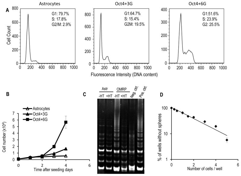
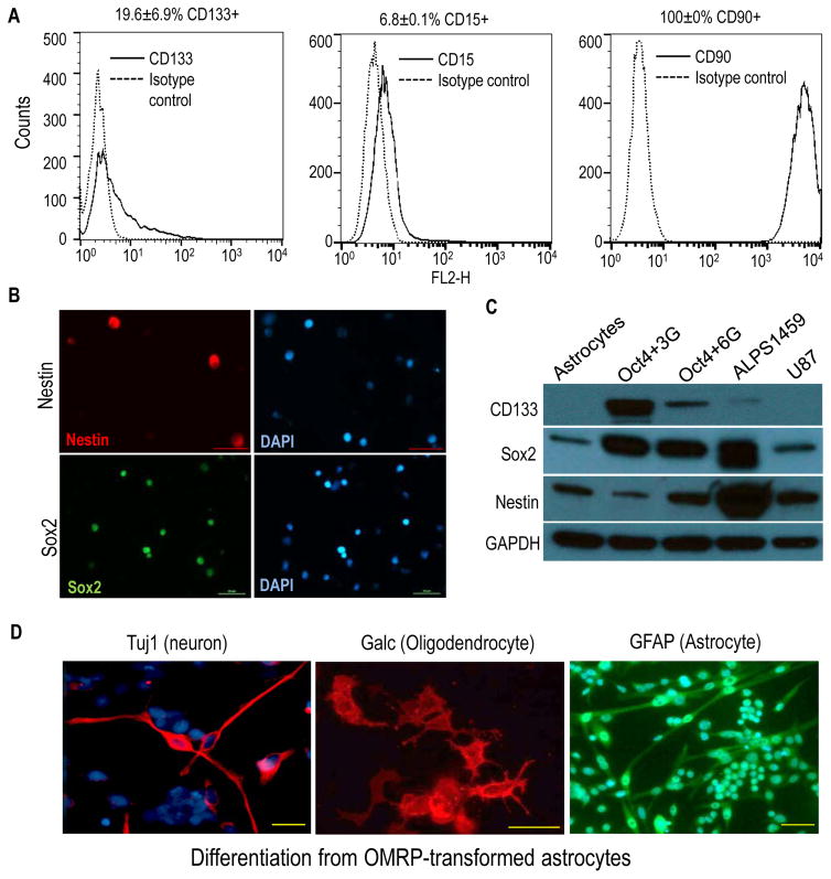
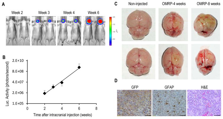
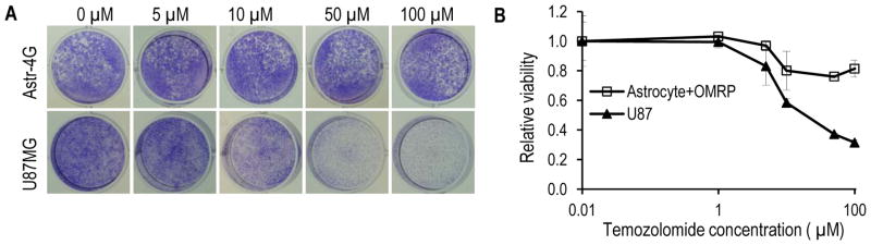
Cancer stem-like cells (CSC) are thought to drive brain cancer, but their cellular and molecular origins remain uncertain. Here, we report the successful generation of induced CSC (iCSC) from primary human astrocytes through the expression of defined genetic factors. Combined transduction of four factors, Myc, Oct-4, p53DD, and Ras, induced efficient transformation of primary human astrocytes into malignant cells with powerful tumor-initiating capabilities. Notably, transplantation of 100 transduced cells into nude mice was sufficient for tumor formation. The cells showed unlimited self-renewal ability with robust telomerase activities. In addition, they expressed typical glioma stem-like cell markers, such as CD133, CD15, and CD90. Moreover, these cells could form spheres in culture and differentiate into neuron-like, astrocyte-like, and oligodendrocyte-like cells. Finally, they also displayed resistance to the widely used brain cancer drug temozolomide. These iCSCs could provide important tools for studies of glioma biology and therapeutics development. Cancer Res; 76(17); 5143-50. ©2016 AACR.


Conflict of interest statement

The authors declare no financial interest in the work described in this study.

Figures

Figure 1
Figure 1. Rapid induction of transformation in primary human astrocytes
A). Changes in astrocyte cellular morphology during the course of induced transformation. Shown are examples from Oct+6G transformed group. The scale bars represent 200 μm. B). Colony outgrowth in astrocytes transformed with a variety of gene combinations, n=3. C). Soft agar colony growth from cells transduced with a variety of gene combinations, n=3. D). Soft agar colony growth from astrocytes transduced with either the Oct4+3G or the Oct4+6G combinations, n=3. In B–D, the error bars represent standard error of the mean (SEM).
Figure 2
Figure 2. Robust self-renewal and sphere-forming abilities of the transformed astrocytes
A). Flow cytometry analysis of the cell cycle distribution profiles of control (primary astrocytes), Oct4+3G− and Oct4+6G− transformed astrocytes. B). Growth curve of control human astrocytes, Oct4+3G− and Oct4+6G-transformed astrocyte cells. C). TRAP assay for telomerase activities of primary astrocytes and OMRP (4G)-transduced astrocytes. HT: heat treatment. Positive and negative controls were provided by manufacturer. D). Results of limited dilution assay for the sphere-forming ability of OMRP-transduced cells. The error bars in B & D represent standard error of the mean.
Figure 3
Figure 3. OMRP-transduced cells exhibit characteristics of cancer stem cells
A). Flow cytometry analysis of cell surface expression of cancer stem cell markers CD133 (left panel), CD15 (mid panel), and CD90 (right panel) in OMRP-transformed cells. B). Immunofluorescence staining of Nestin and Sox2 in OMRP-transduced cells. Scale bars represent 50 μm. C). Western blot analysis of stem cell markers CD133, Sox2, and Nestin in control astrocytes, Oct4+3G gene- and Oct4+7G-gene transformed astrocytes, ALPS1459 (a patient-derived glioma stem cell line), and U87MG, an established glioma cell line. D). Differentiation of OMRP-transformed cells in neuronal, oligodendrocyte, and astrocyte differentiation media. Immunofluorescence staining of characteristic markers (Tuj1, Galc, and GFAP) were done for each of the cell types, respectively. Scale bars represent 50 μm.
Figure 4
Figure 4. Tumor initiation in vivo from OMRP-transduced astrocytes
A). Tumor formation in nude mice from 500 firefly luciferase-labeled, OMRP-transformed astrocytes injected intracranially. Bioluminescence imaging was used to follow tumor growth non-invasively at different time points. B). Exponential increase in intracranial bioluminescence signals from the OMRP-transformed cells during week 2–6. The error bars represent standard error of the mean (SEM), n=5. C). Brains of non-injected mice (left panels) and those injected with OMRP-transformed cells (middle and right panels). Notice with smaller, deformed shapes of the brains and abnormal tissue growth with greenish tint (from EGFP-labeled tumor cells) in the latter group. D). Immunohistochemical (left and middle panels) and H&E (right panel) staining of brain sections from the tumor-bearing mice. Scale bars represent 50 μm.
Fig. 5
Fig. 5
Drug resistance of OMRP-transduced astrocytes. A). Crystal violet staining of OMRP-transformed astrocytes and U87MG cells after treatment with various concentrations of temozolomide. B). Quantitative estimate of the relative survival of OMRP-transduced astrocytes and U87MG cells after temozolomide treatment. The error bars represent standard deviation (n=3).

Similar articles

Cited by

References

    1. Bonnet D, Dick JE. Human acute myeloid leukemia is organized as a hierarchy that originates from a primitive hematopoietic cell. Nat Med. 1997;3:730–7. - PubMed
    1. Al-Hajj M, Wicha MS, Benito-Hernandez A, Morrison SJ, Clarke MF. Prospective identification of tumorigenic breast cancer cells. Proc Natl Acad Sci U S A. 2003;100:3983–8. - PMC - PubMed
    1. Singh SK, Hawkins C, Clarke ID, Squire JA, Bayani J, Hide T, et al. Identification of human brain tumour initiating cells. Nature. 2004;432:396–401. - PubMed
    1. Driessens G, Beck B, Caauwe A, Simons BD, Blanpain C. Defining the mode of tumour growth by clonal analysis. Nature. 2012;488:527–30. - PMC - PubMed
    1. Chen J, Li Y, Yu TS, McKay RM, Burns DK, Kernie SG, et al. A restricted cell population propagates glioblastoma growth after chemotherapy. Nature. 2012;488:522–6. - PMC - PubMed

Publication types