. 2016 Nov;139(4):586-595.
doi: 10.1111/jnc.13720. Epub 2016 Jul 22.

Haptoglobin increases the vulnerability of CD163-expressing neurons to hemoglobin

Affiliations

Haptoglobin increases the vulnerability of CD163-expressing neurons to hemoglobin

Jing Chen-Roetling et al. J Neurochem. 2016 Nov.

Abstract

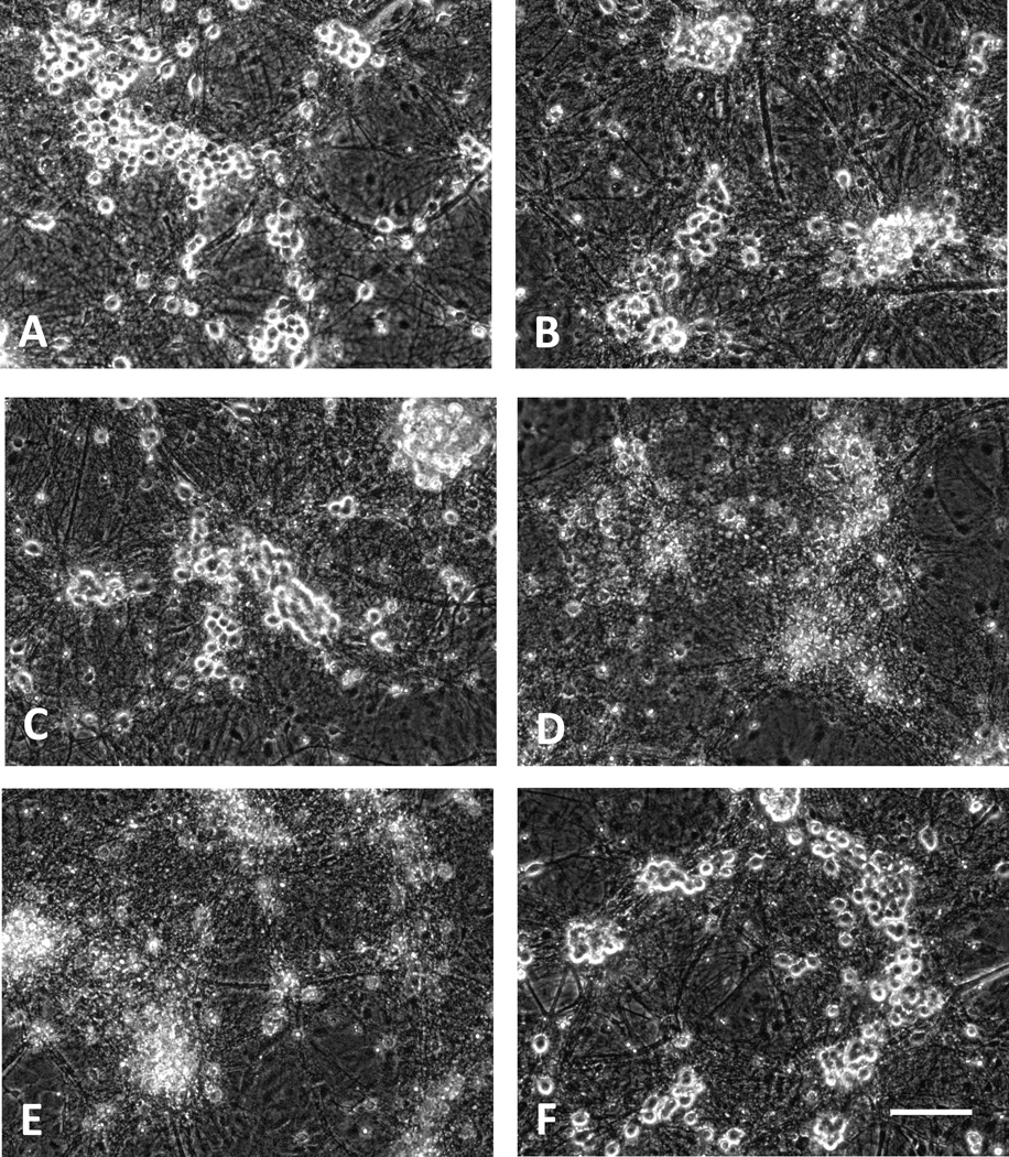
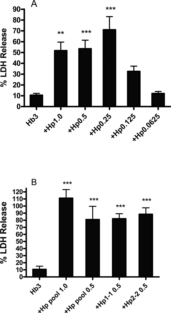
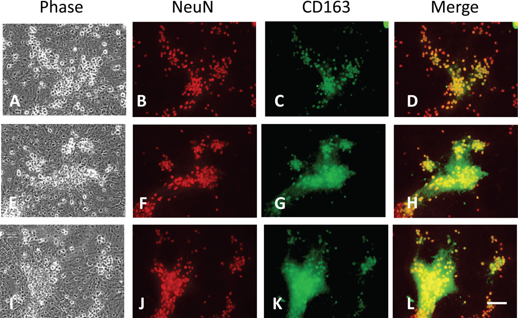
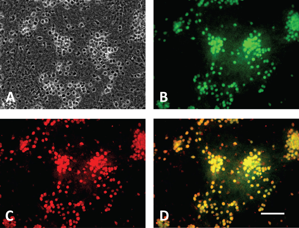
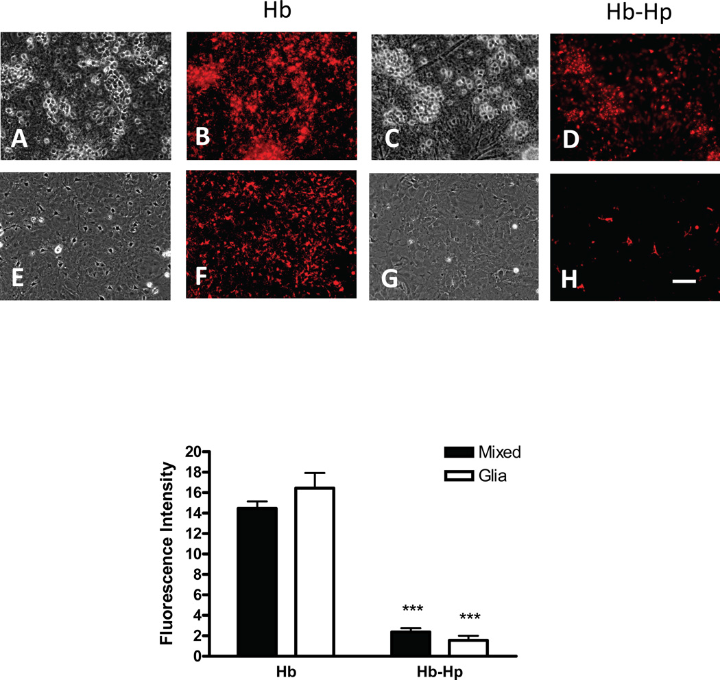
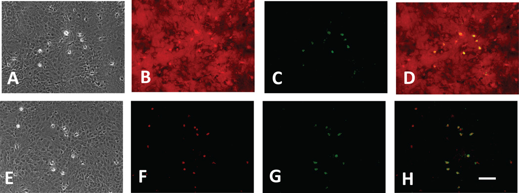
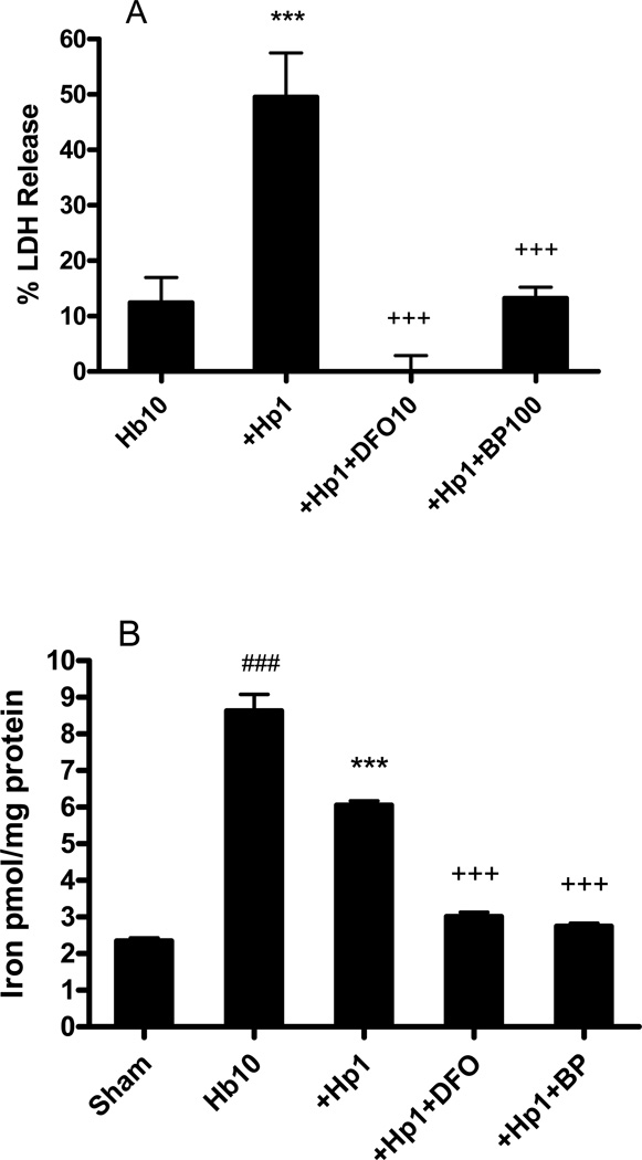
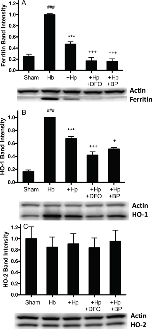
Haptoglobin (Hp) binds hemoglobin (Hb) with high affinity and provides the primary defense against its toxicity after intravascular hemolysis. Neurons are exposed to extracellular Hb after CNS hemorrhage, and a therapeutic effect of Hp via Hb sequestration has been hypothesized. In this study, we tested the hypothesis that Hp protects neurons from Hb in primary mixed cortical cell cultures. Treatment with low micromolar concentrations of human Hb for 24 h resulted in loss of 10-20% of neurons without injuring glia. Concomitant treatment with Hp surprisingly increased neuronal loss five-sevenfold, with similar results produced by Hp 1-1 and 2-2 phenotypes. Consistent with a recent in vivo observation, neurons expressed the CD163 receptor for Hb and the Hb-Hp complex in these cultures. Hp reduced overall Hb uptake, directed it away from the astrocyte-rich CD163-negative glial monolayer, and decreased induction of the iron-binding protein ferritin. Hb-Hp complex neuronal toxicity, like that of Hb per se, was iron-dependent and reduced by deferoxamine and 2,2' bipyridyl. These results suggest that Hp increases the vulnerability of CD163+ neurons to Hb by permitting Hb uptake while attenuating the protective response of ferritin induction by glial cells. Cover Image for this issue: doi: 10.1111/jnc.13342.

Keywords: intracerebral hemorrhage; iron; neurotoxicity; stroke; subarachnoid hemorrhage.


Conflict of interest statement

The authors report no conflicts of interest.

Figures

Figure 1
Figure 1
Morphological appearance of cultures treated with hemoglobin (Hb) and haptoglobin (Hp). A) Phase-contrast image of control culture subjected to medium exchange only. Phase-bright neuron cell bodies are connected by processes and are attached to a confluent monolayer of glial cells. B) Culture treated with 3 µM Hb alone for 24 hours. C–E) Cultures treated with 3 µM Hb plus 0.125 mg/ml, 0.25 mg/ml and 1.0 mg/ml Hp, respectively, demonstrating widespread neuronal loss at higher Hp concentrations. F) Culture treated with 1 mg/ml Hp alone. Scale bar = 100 µm.
Figure 2
Figure 2
Haptoglobin increases neuronal vulnerability to hemoglobin. Percentage neuron loss as measured by LDH release assay in cultures treated for 24 hours with Hb 3µM alone or with indicated concentrations (mg/ml) of haptoglobin. A) Testing BPL haptoglobin, enriched in 1-1 phenotype; B) testing pooled Hp, Hp 1-1 and Hp 2-1 from Athens Research and Technology. The weak LDH signal in control cultures undergoing medium exchange only was subtracted from all values to calculate the signal produced by cell death. All values were scaled to those in sister cultures from the same plating that were treated with 300 µM NMDA (=100), which kills all neurons without injuring glia. **P < 0.01, **P < 0.001 v. value in cultures treated with Hb only, n = 4–12/condition.
Figure 3
Figure 3
Primary cultured neurons express CD163. A–D) Control culture subjected to medium exchange only and fixed 8 hours later, immunostained with antibodies to CD163 and neuronal marker NeuN; CD163 immunoreactivity is largely limited to neuron somata, with minimal staining of neuropil and background glial monolayer. E–H) Culture treated with 10 µM Hb for 8 hours; CD163 immunreactivity has spread to dense neuropil surrounding clusters of neuron somata; I–L) Culture treated with 10 µM Hb plus 1 mg/ml Hp for 8 hours; immunostaining is similar to Hb alone culture, with some loss of phase-bright appearance of somata indicating early neurodegeneration. Scale bar = 100 µm.
Figure 4
Figure 4
Hb-Hp complexes localize to neuron somata. Fixed culture (A, phase contrast optics) was imaged two hours after incubation with anti-NeuN-FITC (B) and Hb-Hp-Alexa Fluor 568 conjugate (C, 1:25 dilution). Merged image (D) demonstrates that most Hb-Hp-Alexa Fluor+ cells are neurons.
Figure 5
Figure 5
Hp reduces Hb uptake. Viable mixed neuron-glial cultures (A–D, fluorescence and corresponding phase contrast images) and glial cultures (E–H) were treated with Hb-Alexa Fluor 568 conjugate (A,B,E,F) or Hb-Hp-Alexa Fluor 568 conjugate (C,D,G,H). Bars represent mean fluorescence intensity of images (n = 5/condition) captured after 8 hour incubation. ***P<0.001 v. corresponding Hb condition. Scale bar = 100 µm.
Figure 6
Figure 6
Hp directs Hb to microglia in glial cultures. Fixed glial cultures were treated with Hb-Alexa Fluor 568 conjugate (A–D) or Hb-Hp-Alexa Fluor 568 conjugate (E–H) for 2 hours. A,E: Phase contrast images showing confluent glial monolayers with scattered small phase-bright cells with the typical appearance of ameboid microglia; B,F) Hb-Alexa Fluor 568 conjugate diffusely stains culture (B), while Hb-Hp-Alexa Fluor 568 conjugate localizes to ameboid cells (F); C,G) Iba-1 immunostaining to detect activated microglia; D,H) Merged images. Scale bar = 100 µm.
Figure 7
Figure 7
Hb-Hp toxicity is iron dependent. A) Percentage neuron loss, as measured by LDH release assay, in cultures treated for 24 h with Hb 10 µM alone, Hb 10 µM plus Hp 1 mg/ml, or Hb, Hp plus indicated concentrations (µM) of deferoxamine (DFO) or 2,2’-bipyridyl (BP). B) Nonheme iron content in cultures treated for 24 hours as in A. ***P < 0.001 v. Hb alone condition, ###P < 0.001 v. sham, +++P < 0.001 v. Hb+Hp, n = 5–13/condition.
Figure 8
Figure 8
Hp reduces ferritin and HO-1 expression in Hb-treated cultures. Ferritin, heme oxygenase-1 (HO-1) and HO-2 expression in cultures treated with Hb 10 µM alone, with Hp 1 mg/ml, or with Hp plus deferoxamine (DFO) 10 µM or 2,2’-Bipyridyl (BP) 100 µM for 8 hours. ***P<0.001 v. Hb alone, ###P<0.001 v. sham, +++P < 0.001, +P < 0.05 v. Hb+Hp, n = 4/condition.

References

    1. Arosio P, Levi S. Ferritin, iron homeostasis, and oxidative damage. Free Radic Biol Med. 2002;33:457–463. - PubMed
    1. ARUP. Haptoglobin: ARUP Lab Tests. 2016
    1. Borsody M, Burke A, Coplin W, Miller-Lotan R, Levy A. Haptoglobin and the development of cerebral artery vasospasm after subarachnoid hemorrhage. Neurology. 2006;66:634–640. - PubMed
    1. Brewer GJ. Serum-free B27/neurobasal medium supports differentiated growth of neurons from the striatum, substantia nigra, septum, cerebral cortex, cerebellum, and dentate gyrus. J Neurosci Res. 1995;42:674–683. - PubMed
    1. Chen-Roetling J, Chen L, Regan RF. Apotransferrin protects cortical neurons from hemoglobin toxicity. Neuropharmacology. 2011;60:423–431. - PMC - PubMed

Publication types

MeSH terms