

Review
doi: 10.1099/jgv.0.000535.
Epub 2016 Jun 30.
Filamentous influenza viruses
Affiliations
- PMID: 27365089
- PMCID: PMC5935222
- DOI: 10.1099/jgv.0.000535
Item in Clipboard
Review
Filamentous influenza viruses
J Gen Virol.
2016 Aug.
Abstract
Clinical isolates of influenza virus produce pleomorphic virus particles, including extremely long filamentous virions. In contrast, strains of influenza that have adapted to laboratory growth typically produce only spherical virions. As a result, the filamentous phenotype has been overlooked in most influenza virus research. Recent advances in imaging and improved animal models have highlighted the distinct structure and functional relevance of filamentous virions. In this review we summarize what is currently known about these strikingly elongated virus particles and discuss their possible roles in clinical infections.
Figures
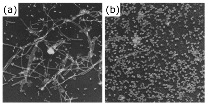
Filamentous influenza virions are clearly visible after two passages of the clinical isolate influenza A/Rockefeller Institute/1/1957 (H2N2) virus in embryonated chicken eggs (a) but are lost following twelve passages (b). Electron micrographs © Choppin et al., 1960. Originally published in THE JOURNAL OF EXPERIMENTAL MEDICINE. 112:945-52.
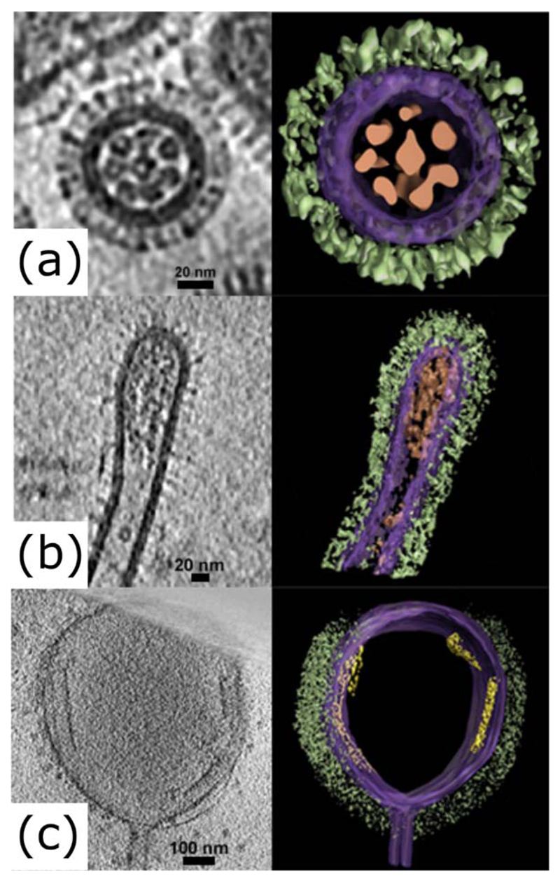
Electron tomograms of influenza virions, showing slices (left panels) and segmented images (right panels) of (a) a transverse section of a bacilliform virion, (b) a longitudinal section of the tip of a filamentous virion and (c) a longitudinal section of an Archetti body at the end of a filamentous virion. Images were manually segmented and coloured to show viral glycoproteins (green), membrane and associated matrix (purple), genome (brown) and putative free M1 sheets (yellow). Tomograms were obtained as part of a previous study (Vijayakrishnan et al., 2013) and manually segmented using Amira (TGS).
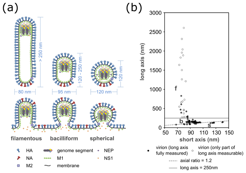
The dimensions of influenza virions, shown (a) as a schematic of budding and released virions, with typical sizes indicated, and (b) as measurements of purified influenza A/Udorn/72 virions. For (a) it should be noted that the incorporation of NS1 and NEP has so far only been examined in spherical virions, and their general incorporation is inferred from this. For (b) measurements of 96 virions were taken by cryoelectron microscopy (data replotted from Vijayakrishnan et al., 2013). Open circles indicate filaments which extended beyond the field of view and so are longer than measured. Spherical virions (s) are distinguished from bacilliform virions (b) by having an axial ratio less than 1.2 (dashed line); filaments (f) are distinguished from bacilliform virions by having a length greater than 250 nm (solid line).
References
-
- Ada GL, Perry BT. Properties of the nucleic acid of the Ryan strain of filamentous influenza virus. Journal of general microbiology. 1958;19:40–54. - PubMed
-
- Ada GL, Perry BT, Abbot A. Biological and physical properties of the Ryan strain of filamentous influenza virus. J Gen Microbiol. 1958;19:23–39. - PubMed
-
- Ada GL, Perry BT, Edney M. Infectivity of influenza virus filaments. Nature. 1957;180:1134. - PubMed
-
- Al-Mubarak F, Daly J, Christie D, Fountain D, Dunham SP. Identification of morphological differences between avian influenza A viruses grown in chicken and duck cells. Virus research. 2015 - PubMed
Publication types
MeSH terms
Grants and funding
LinkOut - more resources
Full Text Sources
Other Literature Sources