. 2016 Sep;5(9):1277-88.
doi: 10.5966/sctm.2015-0223. Epub 2016 Jun 30.

Preischemic Administration of Nonexpanded Adipose Stromal Vascular Fraction Attenuates Acute Renal Ischemia/Reperfusion Injury and Fibrosis

Affiliations

Preischemic Administration of Nonexpanded Adipose Stromal Vascular Fraction Attenuates Acute Renal Ischemia/Reperfusion Injury and Fibrosis

Liuhua Zhou et al. Stem Cells Transl Med. 2016 Sep.

Abstract

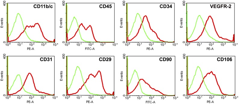
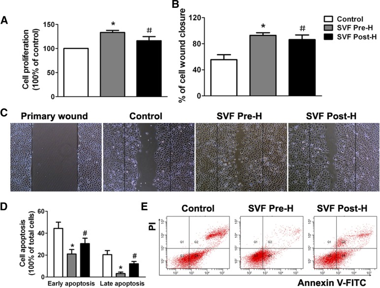
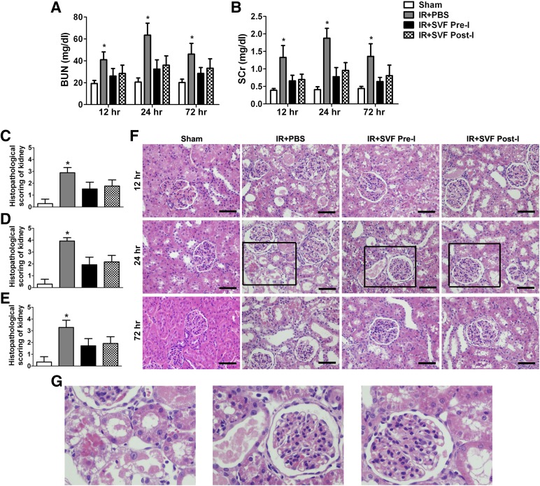
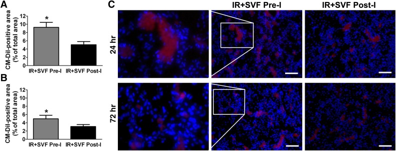
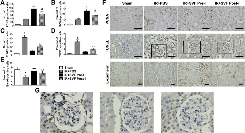
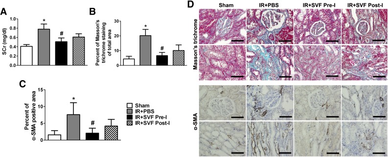
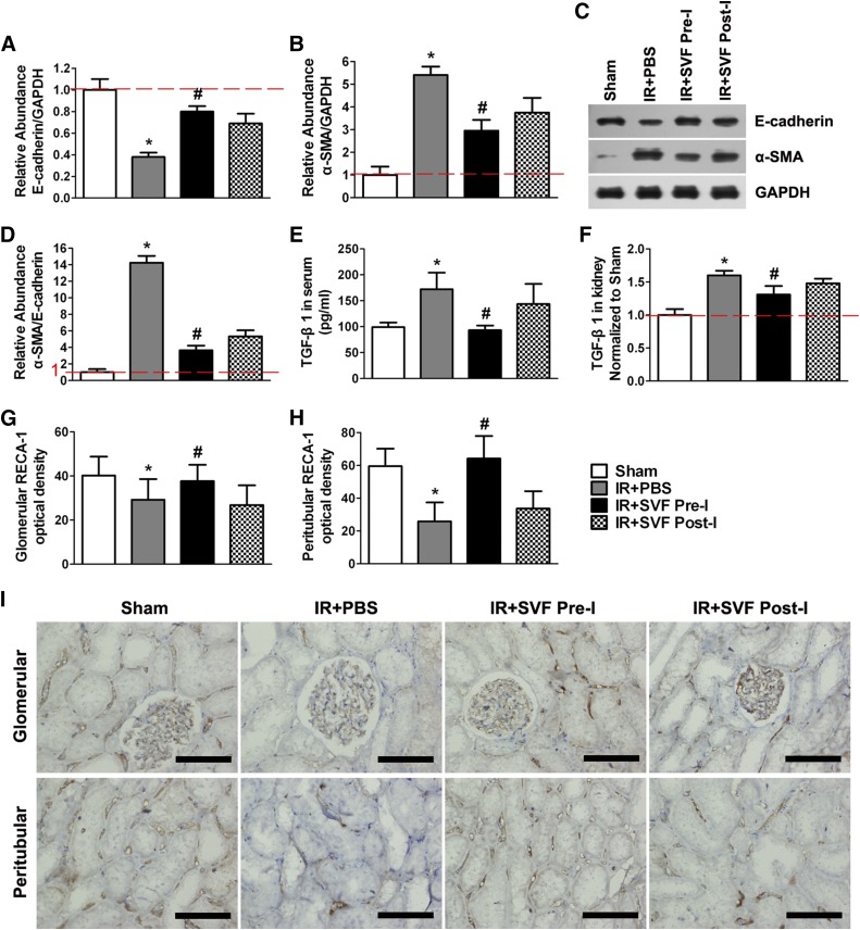
: Ischemia/reperfusion (IR)-induced acute kidney injury (AKI) is a common clinical syndrome. Stem/progenitor cell therapy is a promising option to foster the intrinsic capacity for kidney regeneration. However, there are still several challenges to be resolved, including the potential risks during cell culture, low retention rate after transplantation, and unclear effect on the progression of chronic kidney disease (CKD). Recently, nonexpanded adipose stromal vascular fraction (SVF) has been regarded as an attractive cell source for cell-based therapy. Preconditioning with ischemia has been suggested as a useful method to promote the retention and survival of transplanted cells in vivo. In this study, freshly isolated autologous SVF was transplanted to the kidney of rats before ischemia, and then an IR-induced AKI model was established. Postischemic administration of SVF to the kidney was performed after renal IR injury was induced. A higher cell retention rate was detected in the preischemic group. Preischemic administration of SVF showed stronger functional and morphologic protection from renal IR injury than postischemic administration, through enhancing tubular cell proliferation and reducing apoptosis. Progression of kidney fibrosis was also significantly delayed by preischemic administration of SVF, which exhibited stronger inhibition of transforming growth factor-β1-induced epithelia-mesenchymal transition and microvascular rarefaction. In addition, in vitro study showed that prehypoxic administration of SVF could significantly promote the proliferation, migration, and survival of hypoxic renal tubular epithelial cells. In conclusion, our study demonstrated that preischemic administration of nonexpanded adipose SVF protected the kidney from both acute IR injury and long-term risk of developing CKD.

Significance: Renal ischemia/reperfusion (IR) injury is a common clinical syndrome. Cell-based therapy provides a promising option to promote renal repair after IR injury. However, several challenges still remain because of the potential risks during cell culture, low retention rate after transplantation, and unclear effect on the progression of chronic kidney disease. Stromal vascular fraction (SVF) is considered as an attractive cell source. This study demonstrated that preischemic administration of uncultured SVF could increase cell retention and then improve renal function and structure at both early and long-term stage after IR, which may provide a novel therapeutic approach for IR injury.

Keywords: Adipose stem cells; Autologous stem cell transplantation; Cellular therapy; Ischemia/reperfusion; Renal; Stromal vascular fraction.


Figures

Figure 1.
Figure 1.
Characterization of freshly isolated stromal vascular fraction (SVF) by flow cytometric analysis. Representative flow cytometry histograms of SVF showed that freshly isolated SVF could express hematopoietic, mesenchymal, and endothelial markers. Abbreviations: FITC-A, fluorescein isothiocyanate area; PE-A, phycoerythrin area; VEGFR-2, vascular endothelial growth factor 2.
Figure 2.
Figure 2.
In vitro effects of SVF prehypoxic administration on renal tubular epithelial cell (TEC) in hypoxic environment. (A): Prehypoxic administration of SVF could significantly promote the proliferation of hypoxic renal TEC, compared with the control and posthypoxic administration groups. (B): Prehypoxic administration of SVF could significantly promote the scratch wound healing of hypoxic TEC, compared with the control and posthypoxic administration groups. (C): Representative images of cell scratch wound healing (magnification, ×100). (D): Prehypoxic administration of SVF could significantly reduce the apoptosis of hypoxic TEC, compared with the control and posthypoxic administration groups. (E): Representative flow cytometry histograms of Annexin V/PI apoptosis assay in different groups. ∗, p < .05 (vs. control and SVF post-H); #, p < .05 (vs. control). Abbreviations: FITC, fluorescein isothiocyanate; PI, propidium iodide; SVF, stromal vascular fraction; SVF Post-H, posthypoxic administration of stromal vascular fraction; SVF Pre-H, prehypoxic administration of stromal vascular fraction.
Figure 3.
Figure 3.
Early protective effect of stromal vascular fraction (SVF) preischemic administration on acute renal IR injury. (A, B): Rats that received SVF administration showed significantly lower BUN (A) and SCr (B) values at 12, 24, and 72 hours after IR compared with control group. (C–E): SVF administration could also significantly reduce tubular injury score of kidneys in rats at 12 (C), 24 (D), and 72 (E) hours after IR compared with the control group. Although there was no significant difference in renal function and histopathological score between preischemic and postischemic groups, preischemic administration of SVF still contributed to lower BUN and SCr values, as well as reduced tubular injury, compared with postischemic administration. (F, G): Representative images of H&E staining preformed on sections of kidneys at 12, 24, and 72 hours after IR in different groups (magnification, ×400). Scale bars = 50 μm. ∗, p < .05 (vs. sham, IR+SVF pre-I, and IR+SVF post-I). Abbreviations: BUN, blood urea nitrogen; IR, ischemia/reperfusion; PBS, phosphate-buffered saline; SCr, serum creatinine; SVF Post-I, postischemic administration of stromal vascular fraction; SVF Pre-I, preischemic administration of stromal vascular fraction.
Figure 4.
Figure 4.
Cell tracking in rats treated with stromal vascular fraction (SVF). (A, B): Percentages of CM-DiI-positive areas in kidney sections of rats at 24 (A) and 72 (B) hours after IR were calculated. CM-DiI-positive SVF was significantly increased in the kidneys that received preischemic administration of SVF compared with those that underwent postischemic treatment. (C): Representative images of CM-DiI-labeled SVF in the kidneys at 24 and 72 hours after IR at different groups (magnification, ×200). Scale bars = 100 μm. ∗, p < .05 (vs. IR+SVF post-I). Abbreviations: IR, ischemia/reperfusion; SVF Post-I, postischemic administration of stromal vascular fraction; SVF Pre-I, preischemic administration of stromal vascular fraction.
Figure 5.
Figure 5.
Immunohistochemical staining of PCNA, E-cadherin, and TUNEL staining in kidney sections at 72 hours after IR in different groups. (A–E): Quantitative analysis of PCNA-positive cells (A, B), TUNEL positive cells (C, D), and E-cadherin-positive cells (E) was performed by using Image-Pro Plus software. (F, G): Representative images of PCNA, E-cadherin, and TUNEL staining in the kidneys at 72 hours after IR at different groups (magnification, ×400 [PCNA and TUNEL staining], ×200 [E-cadherin staining]). Scale bars = 50 μm. ∗, p < .05 (vs. sham, IR+PBS, and IR+SVF post-I); #, p < .05 (vs. IR+PBS); Δ, p < .05 (vs. sham, IR+SVF pre-I, and IR+SVF post-I); ∗∗, p < .05 (vs. IR+SVF pre-I). Abbreviations: E-cad, E-cadherin; IR, ischemia/reperfurion; PBS, phosphate-buffered saline; PCNA, proliferating cell nuclear antigen; SVF Post-I, postischemic administration of stromal vascular fraction; SVF Pre-I, preischemic administration of stromal vascular fraction; TUNEL, terminal transferase-mediated deoxyuridine triphosphate nick-end-labeling.
Figure 6.
Figure 6.
Long-term preservation of renal function and inhibition of fibrosis in tubulointerstitial areas by preischemic administration of stromal vascular fraction (SVF). (A): SCr value at 6 months after IR in different groups. (B, C): Quantitative analysis of Masson’s trichrome staining (B) and α-SMA positive area (C) was performed by using Image-Pro Plus software. (D): Representative images of Masson’s trichrome and α-SMA staining in the kidneys at 6 months after IR at different groups (magnification, ×400). Scale bars = 50 μm. ∗, p < .05 (vs. sham, IR+SVF pre-I, and IR+SVF post-I); #, p < .05 (vs. IR+SVF post-I). Abbreviations: IR, ischemia/reperfusion; SVF, stromal vascular fraction; PBS, phosphate-buffered saline; SCr, serum creatinine; α-SMA, α-smooth muscle actin; SVF Post-I, postischemic administration of stromal vascular fraction; SVF Pre-I, preischemic administration of stromal vascular fraction.
Figure 7.
Figure 7.
Preischemic administration of stromal vascular fraction (SVF) inhibited TGF-β1-induced epithelia-mesenchymal transition (EMT) and microvascular rarefaction. (A–C): Relative abundance of E-cadherin/GAPDH (A) and α-SMA/GAPDH (B) was calculated by Western blot assay (C) at 6 months after IR in different groups. (D): Preischemic administration of SVF significantly attenuated the upregulation of α-SMA/E-cadherin expression compared with postischemic administration of SVF or PBS (p < .05). (E, F): The expression levels of TGF-β1 in serum (E) and injured kidney (F) in rats received preischemic administration of SVF were significantly lower than that received postischemic administration of SVF or PBS. (G, H): Mean optical density was evaluated by RECA-1 staining in glomerular (G) and peritubular (H) areas in kidney sections of rats at 6 months after IR. Preischemic administration of SVF significantly inhibited microvascular rarefaction in kidney sections compared with postischemic administration. (I): Representative microscopic images of RECA-1 staining preformed on sections of kidneys at 6 months after IR in different groups (×400). Scale bars = 50 μm. ∗, p < .05 (vs. sham, IR+SVF pre-I, and IR+SVF post-I); #, p < .05 (vs. IR+SVF post-I). Abbreviations: GAPDH, glyceraldehyde-3-phosphate dehydrogenase; IR, ischemia/reperfusion; PBS, phosphate-buffered saline; RECA-1, rat endothelial cell antigen-1; α-SMA, α-smooth muscle actin; SVF Post-I, postischemic administration of stromal vascular fraction; SVF Pre-I, preischemic administration of stromal vascular fraction; TGF-β1, transforming growth factor-β1.

References

    1. Liaño F, Pascual J, Madrid Acute Renal Failure Study Group Epidemiology of acute renal failure: A prospective, multicenter, community-based study. Kidney Int. 1996;50:811–818. - PubMed
    1. Bonventre JV, Yang L. Cellular pathophysiology of ischemic acute kidney injury. J Clin Invest. 2011;121:4210–4221. - PMC - PubMed
    1. Sharfuddin AA, Molitoris BA. Pathophysiology of ischemic acute kidney injury. Nat Rev Nephrol. 2011;7:189–200. - PubMed
    1. Leung KC, Tonelli M, James MT. Chronic kidney disease following acute kidney injury-risk and outcomes. Nat Rev Nephrol. 2013;9:77–85. - PubMed
    1. Iwano M, Plieth D, Danoff TM, et al. Evidence that fibroblasts derive from epithelium during tissue fibrosis. J Clin Invest. 2002;110:341–350. - PMC - PubMed