. 2016 Dec;100(6):1335-1347.
doi: 10.1189/jlb.2A1215-555RR. Epub 2016 Jun 30.

FcγR mediates TLR2- and Syk-dependent NLRP3 inflammasome activation by inactivated Francisella tularensis LVS immune complexes

Affiliations

FcγR mediates TLR2- and Syk-dependent NLRP3 inflammasome activation by inactivated Francisella tularensis LVS immune complexes

Ellen B Duffy et al. J Leukoc Biol. 2016 Dec.

Abstract

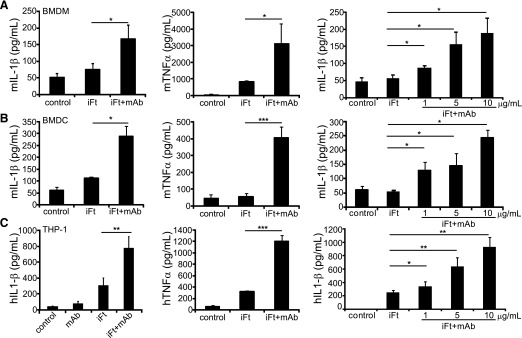
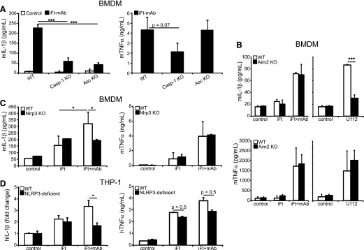
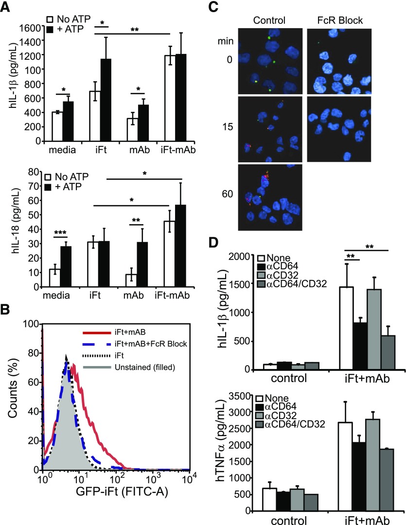
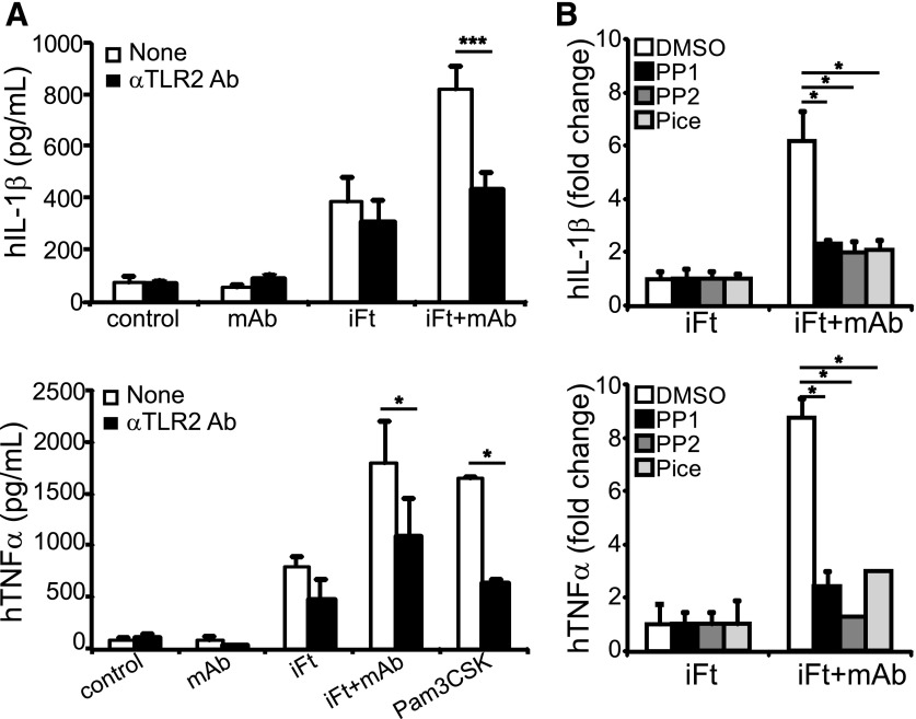
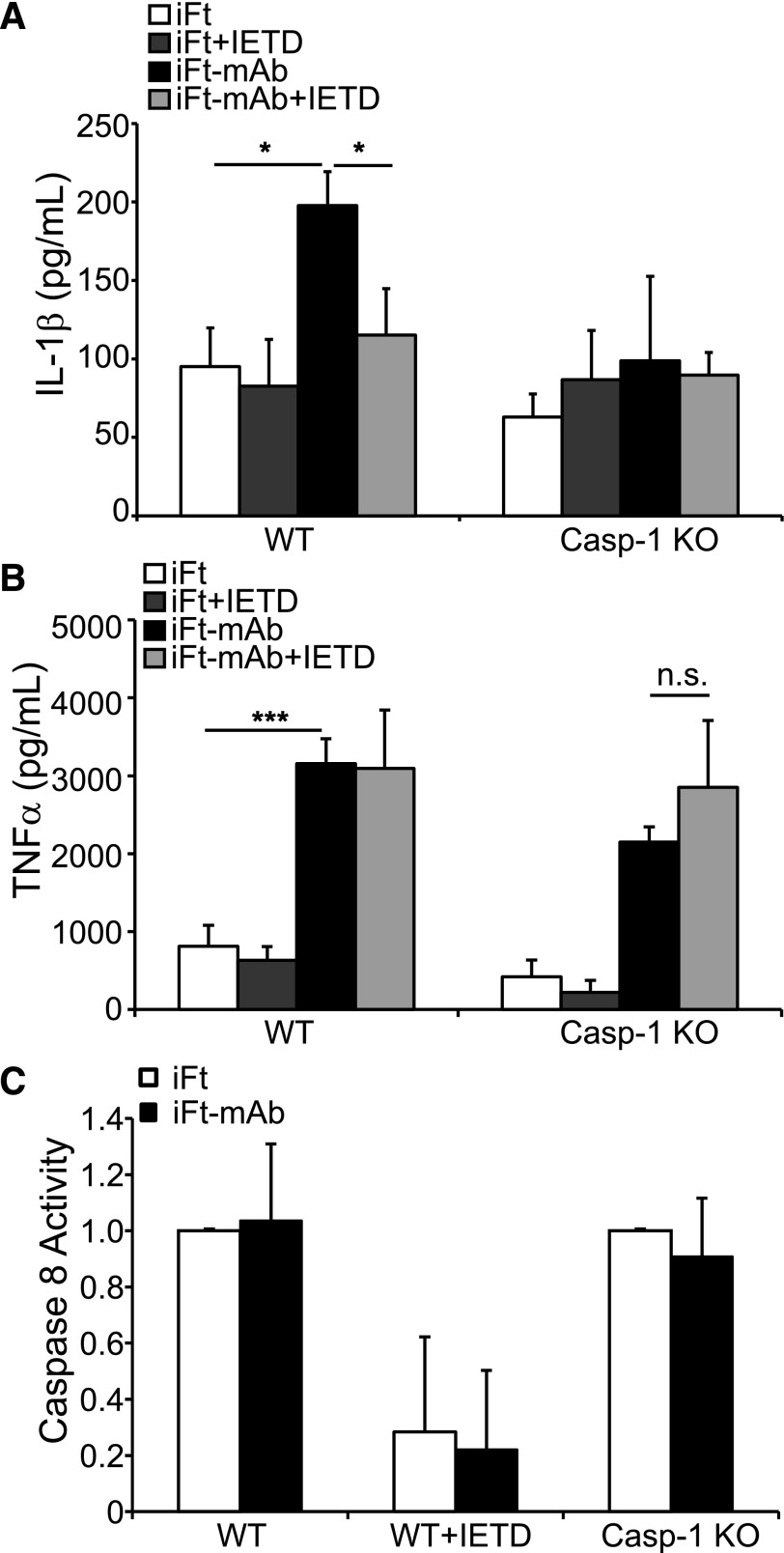
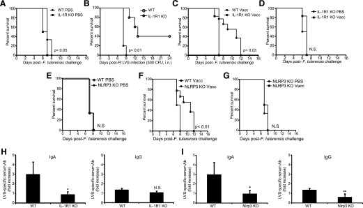
IgG (mAb)-opsonized, inactivated Francisella tularensis LVS (iFt-mAb) enhances TLR2-dependent IL-6 production by macrophages via Fcγ receptors (FcγR). In mice, vaccination with iFt-mAb provides IgA-dependent protection against lethal challenge with Ft LVS. Because inflammasome maturation of IL-1β is thought important for antibody-mediated immunity, we considered the possibility that iFt-mAb elicits an FcγR-dependent myeloid cell inflammasome response. Herein, we find that iFt-mAb enhances macrophage and dendritic cell IL-1β responses in a TLR2- and FcγR-dependent fashion. Although iFt-mAb complexes bind FcγR and are internalized, sensing of cytosolic DNA by absent in melanoma 2 (AIM2) is not required for the IL-1β response. In contrast, ASC, caspase-1, and NLR family pyrin domain-containing 3 (NLRP3) are indispensable. Further, FcγR-mediated spleen tyrosine kinase (Syk) signaling is required for this NLRP3-dependent IL-1β response, but the alternative IL-1β convertase caspase-8 is insufficient. Finally, iFt-mAb-vaccinated wild-type mice exhibit a significant delay in time to death, but IL-1R1- or Nlrp3-deficient mice vaccinated in this way are not protected and lack appreciable Francisella-specific antibodies. This study demonstrates that FcγR-mediated Syk activation leads to NLRP3 inflammasome-dependent IL-1β production in macrophages and suggests that an Nlrp3- and IL-1R-dependent process contributes to the IgA response important for protection against Ft LVS. These findings extend our understanding of cellular responses to inactivated pathogen-opsonized vaccine, establish FcγR-elicited Syk kinase-mediated NLRP3 inflammasome activation, and provide additional insight toward understanding crosstalk between TLR and FcγR signals.

Keywords: IL-1β; macrophage; vaccine.


Figures

Figure 1.
Figure 1.. The iFt-mAb complex activates cytokine production in macrophages and BMDCs.
BMDM (A) and BMDC (B) from WT mice or PMA-treated THP-1 (C) were left untreated (media control) or stimulated for 24 h with iFt, mAb, or iFt-mAb at 5 µg/ml or at increasing concentrations. IL-1β and TNF-α were analyzed by ELISA using cell-free supernatants in duplicate. BMDM and THP-1 stimulations are means and sem of 3 independent experiments. BMDC results are means and sd of 2 independent experiments. Student’s t test was used to compare iFt to iFt-mAb. *P < 0.05; **P < 0.01; ***P < 0.001.
Figure 2.
Figure 2.. The NLRP3 inflammasome is required for IL-1β production by iFt-mAb.
BMDM from WT, Casp-1 KO, or Asc KO (A), Aim2 KO (B), and Nlrp3 KO (C) mice were left untreated (media control) or stimulated with iFt or iFt-mAb or infected with Fn U112 (100 MOI) for 24 h before collection of cell-free culture supernatants for IL-1β and TNF-α ELISA. IL-1β and TNF-α analyzed by ELISA of THP-1– and NLRP3-deficient THP-1 24 h poststimulation with iFt or iFt-mAb. Control concentrations of IL-1β (pg/ml) for THP-1– and NLRP3-deficient THP-1 were 275.2 (sd 120.7) and 21.2 (sd 3.1), respectively (D). Results are means of 3 independent experiments with error bars indicating sem. *P < 0.05; ***P < 0.001.
Figure 3.
Figure 3.. FcγR mediates inflammasome activation to enhance IL-1β.
(A) THP-1 were stimulated with iFt, mAb, or iFt-mAb for 23.5 h followed by treatment for 30 min with ATP (5 mM) before collection of cell-free culture supernatants for IL-1β or IL-18 by multiplex immunoassay. (B) Binding of GFP-iFt-mAb to THP-1 with or without FcγR blocking Abs as indicated by flow cytometry counting of cells labeled with GFP-iFt-mAb. The 1-dimensional histogram is 1 of 3 independent experiments with similar results. The filled curve is unstained cells, the solid curve is GFP-iFt-mAb without blocking Abs, the dashed curved is GFP-iFt-mAb with blocking Abs, and the dotted curve is GFP-iFt without mAb and without blocking Abs. THP-1 stimulated with iFt-mAb with and without FcγR blocking Abs were stained with Abs against mAbs (C). Green fluorescence indicates extracellular iFt-mAb; red fluorescence indicates intracellular iFt-mAb. Images were obtained by confocal microscopy. (D) Images are representative of 3 independent experiments with similar results. IL-1β and TNF-α from cell-free supernatants of THP-1 in the presence or absence of anti-FcγR blocking Abs. Shown are means of 3 independent experiments with error bars indicating sem. *P < 0.05; **P < 0.01; ***P < 0.001.
Figure 4.
Figure 4.. TLR2 and Src/Syk signaling are important for cytokine production with iFt-mAb.
IL-1β and TNF-α from cell-free supernatants of THP-1 stimulated for 24 h in the presence or absence of anti-TLR2 blocking Ab (A), with or without 30 min pretreatment with Syk (Pice, piceattenol, 5 µM) and Src inhibitors (PP1 and PP2, 10 μM) (B). Shown are means of 3 independent experiments with error bars indicating sem, except TNF-α in (B), which is the means of 2 independent experiments with triplicate samples and error bars indicating sd. *P < 0.05.
Figure 5.
Figure 5.. Caspase-8 activity is necessary but insufficient for FcγR-mediated IL-1β.
BMDM from WT or Casp-1 KO mice were stimulated for 24 h with iFt or iFt-mAb, with or without pretreatment with a caspase-8 inhibitor (IETD), and IL-1β (A) and TNF-α (B) were analyzed by ELISA in cell-free supernatants. Caspase-8 activity in BMDM from WT or Casp-1 KO mice assayed using a luminescence detection system (C). Data shown reflect the means and sd of triplicate samples and are representative of 3 independent experiments with similar results. *P < 0.05; n.s., not significant.
Figure 6.
Figure 6.. IL-1 and NLRP3 are important for survival against respiratory Ft-LVS in mice.
Groups of 10 WT, IL-1r1 KO, or Nlrp3 KO mice were immunized i.n. with either 20 μl of vehicle (PBS) or 20 μl of 2 × 10⁷ inactivated organisms complexed with 1 μg/ml mAb (iFt-mAb) in PBS (Vacc). Mice were immunized on d 0 and d 21 and then challenged i.n. on d 35 with 20,000 CFU live Ft-LVS and monitored for survival (A, C, D, E, F, and G). Survival curves in all 6 of these panels are from the same groups of mice and are separated for ease of comparison. Results from 1 of 2 vaccination studies with similar results are shown. Groups of 6 WT or IL-1R1 KO mice were infected i.n. with 500 CFU Ft LVS and observed for survival (B). Serum from WT and IL-1r1 mice was collected before challenge, and relative Ft LVS-specific Ab levels for IgA and IgG were measured by ELISA (H). Serum from WT and Nlrp3 mice was collected before challenge, and relative Ft LVS-specific Ab levels for IgA and IgG were measured by ELISA (I). The bars represent the average of Ft LVS-specific Ab detected in serum of iFt-mAb treated mice normalized to the corresponding average unvaccinated serum Ab level. Survival curves were compared using the log-rank test (Mantel-Cox). *P < 0.05; N.S., not significant.

References

    1. Allen I. C., Scull M. A., Moore C. B., Holl E. K., McElvania-TeKippe E., Taxman D. J., Guthrie E. H., Pickles R. J., Ting J. P. (2009) The NLRP3 inflammasome mediates in vivo innate immunity to influenza A virus through recognition of viral RNA. Immunity 30, 556–565. - PMC - PubMed
    1. Gross O., Poeck H., Bscheider M., Dostert C., Hannesschläger N., Endres S., Hartmann G., Tardivel A., Schweighoffer E., Tybulewicz V., Mocsai A., Tschopp J., Ruland J. (2009) Syk kinase signalling couples to the Nlrp3 inflammasome for anti-fungal host defence. Nature 459, 433–436. - PubMed
    1. He X., Mekasha S., Mavrogiorgos N., Fitzgerald K. A., Lien E., Ingalls R. R. (2010) Inflammation and fibrosis during Chlamydia pneumoniae infection is regulated by IL-1 and the NLRP3/ASC inflammasome. J. Immunol. 184, 5743–5754. - PMC - PubMed
    1. Schmitz N., Kurrer M., Bachmann M. F., Kopf M. (2005) Interleukin-1 is responsible for acute lung immunopathology but increases survival of respiratory influenza virus infection. J. Virol. 79, 6441–6448. - PMC - PubMed
    1. Eisenbarth S. C., Colegio O. R., O’Connor W., Sutterwala F. S., Flavell R. A. (2008) Crucial role for the Nalp3 inflammasome in the immunostimulatory properties of aluminium adjuvants. Nature 453, 1122–1126. - PMC - PubMed

MeSH terms