

Intracavitary Irradiation as a Safe Alternative for Cystic Craniopharyngiomas: Case Report and Review of the Literature
- PMID: 27366151
- PMCID: PMC4912999
- DOI: 10.1155/2016/3601395
Intracavitary Irradiation as a Safe Alternative for Cystic Craniopharyngiomas: Case Report and Review of the Literature
Abstract
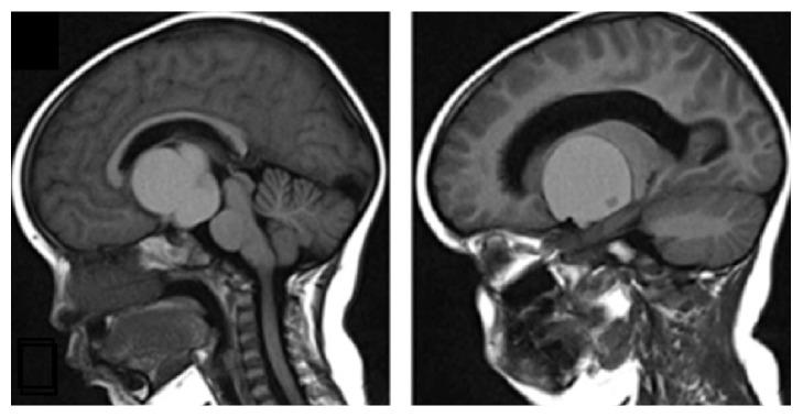
Craniopharyngioma treatment remains a challenge for clinicians and patients. There are many treatment alternatives; however one of them (intracavitary irradiation) seeks to control this type of benign brain tumor using minimally invasive techniques, with the specific aim of avoiding causing significant damage to important structures surrounding the sellar/suprasellar region. We present the case of a 3-year-old patient with a predominantly cystic craniopharyngioma who underwent intracavitary irradiation by stereotactic placement. Using this approach, the patient showed a successful response with remission of headaches and hydrocephalus. A reduction in the size of the cyst was achieved, without deterioration of visual fields, with no hormonal supplementation being needed, and with no evidence of focal neurological signs.
Figures


References
-
- Bülow B., Attewell R., Hagmar L., Malmström P., Nordström C.-H., Erfurth E. M. Postoperative prognosis in craniopharyngioma with respect to cardiovascular mortality, survival, and tumor recurrence. Journal of Clinical Endocrinology and Metabolism. 1998;83(11):3897–3904. - PubMed
LinkOut - more resources
Full Text Sources
Other Literature Sources
