

Brainstem epidermoid cyst: An update
- PMID: 27366244
- PMCID: PMC4849286
- DOI: 10.4103/1793-5482.145163
Brainstem epidermoid cyst: An update
Abstract
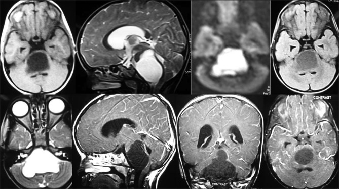
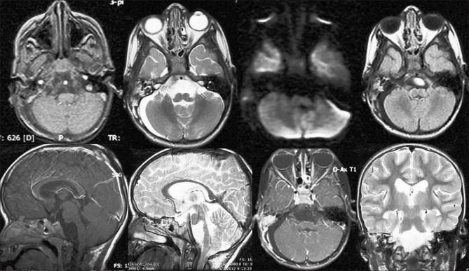
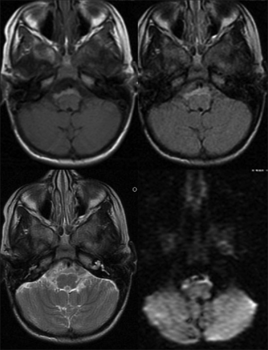
The incidence of epidermoid tumors is between 1% and 2% of all intracranial tumors. The usual locations of epidermoid tumor are the parasellar region and cerebellopontine angle, and it is less commonly located in sylvian fissure, suprasellar region, cerebral and cerebellar hemispheres, and lateral and fourth ventricles. Epidermoid cysts located in the posterior fossa usually arise in the lateral subarachnoid cisterns, and those located in the brain stem are rare. These epidermoids contain cheesy and flaky white soft putty like contents. Epidermoid cysts are very slow growing tumors having a similar growth pattern of the epidermal cells of the skin and develop from remnants of epidermal elements during closure of the neural groove and disjunction of the surface ectoderm with neural ectoderm between the third and fifth weeks of embryonic life. We are presenting an interesting case of intrinsic brainstem epidermoid cyst containing milky white liquefied material with flakes in a 5-year-old girl. Diffusion-weighted imaging is definitive for the diagnosis. Ideal treatment of choice is removal of cystic components with complete resection of capsule. Although radical resection will prevent recurrence, in view of very thin firmly adherent capsule to brainstem, it is not always possible to do complete resection of capsule without any neurological deficits.
Keywords: Brainstem; dermoid; diffusion-weighted imaging; epidermoid; epidermoid cyst; intrinsic; pons.
Conflict of interest statement
Figures
References
-
- Cruveilhier J. Paris: Baillere; 1829. Anatomie pathologique du corps humain. Vol 1, book 2.
-
- Caldarelli M, Colosimo C, Di Rocco C. Intra-axial dermoid/epidermoid tumors of the brainstem in children. Surg Neurol. 2001;56:97–105. - PubMed
-
- Caldarelli M, Massimi L, Kondageski C, Di Rocco C. Intracranial midline dermoid and epidermoid cysts in children. J Neurosurg. 2004;100:473–80. - PubMed
-
- Guidetti B, Gagliardi FM. Epidermoid and dermoid cysts: Clinical evaluation and late surgical results. J Neurosurg. 1977;47:12–8. - PubMed
-
- Kachhara R, Bhattacharya RN, Radhakrishnan VV. Epidermoid cyst involving the brain stem. Acta Neurochir (Wien) 2000;142:97–100. - PubMed
Publication types
LinkOut - more resources
Full Text Sources
Other Literature Sources