Good outcome after delayed surgery for orbitocranial non-missile penetrating brain injury
- PMID: 27366265
- PMCID: PMC4849307
- DOI: 10.4103/1793-5482.179641
Good outcome after delayed surgery for orbitocranial non-missile penetrating brain injury
Abstract
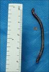
Nonmissile orbitocranial penetrating brain injuries are uncommonly dealt with in a civilian context. Surgical management is controversial, due to the lack of widely accepted guidelines. A 52-year-old man was hit in his left eye by a metallic foreign body (FB). Head computed tomography (CT) scan showed a left subcortical parietal FB with a considerable hemorrhagic trail originating from the left orbital roof. Surgical treatment was staged; an exenteratio oculi and a left parietal craniotomy to extract the FB under intraoperative CT guidance were performed at post trauma day third and sixth, respectively. A postoperative infectious complication was treated conservatively. The patient retained a right hemiparesis (3/5) and was transferred to rehabilitation in good clinical conditions at day 49(th). He had suspended antiepilectic therapy at that time. A case-by-case tailored approach is mandatory to achieve the best outcome in such a heterogeneous nosological entity. Case reporting is crucial to further understand its mechanism and dynamics.
Keywords: Brain abscess; foreign body; intracranial; orbitocranial; penetrating brain injury.
Figures


Similar articles
-
Trans-orbital orbitocranial penetrating injury by pointed iron rod.J Neurosci Rural Pract. 2015 Apr-Jun;6(2):231-3. doi: 10.4103/0976-3147.150282. J Neurosci Rural Pract. 2015. PMID: 25883487 Free PMC article.
-
Nonmissile Penetrating Injury to the Head: Experience with 17 Cases.World Neurosurg. 2016 Oct;94:529-543. doi: 10.1016/j.wneu.2016.06.062. Epub 2016 Jun 24. World Neurosurg. 2016. PMID: 27350299
-
Penetrating orbitocranial gunshot injuries.Surg Neurol. 2005 Jan;63(1):24-30; discussion 31. doi: 10.1016/j.surneu.2004.05.043. Surg Neurol. 2005. PMID: 15639513
-
Treatment of Penetrating Nonmissile Traumatic Brain Injury. Case Series and Review of the Literature.World Neurosurg. 2016 Jul;91:297-307. doi: 10.1016/j.wneu.2016.04.012. Epub 2016 Apr 9. World Neurosurg. 2016. PMID: 27072332 Review.
-
SURGICAL MANAGEMENT OF A PENETRATING BRAIN WOUND AND ASSOCIATED PERFORATING OCULAR INJURY CAUSED BY A LOW-VELOCITY SHARP METALLIC OBJECT: A CASE REPORT AND LITERATURE REVIEW.Acta Clin Croat. 2022 Nov;61(3):537-546. doi: 10.20471/acc.2022.61.03.21. Acta Clin Croat. 2022. PMID: 37492370 Free PMC article. Review.
Cited by
-
Transorbital nonmissile penetrating brain injury: Report of two cases.World J Clin Cases. 2020 Jan 26;8(2):471-478. doi: 10.12998/wjcc.v8.i2.471. World J Clin Cases. 2020. PMID: 32047800 Free PMC article.
References
-
- Chibbaro S, Tacconi L. Orbito-cranial injuries caused by penetrating non-missile foreign bodies. Experience with eighteen patients. Acta Neurochir (Wien) 2006;148:937–41. - PubMed
-
- Abdoli A, Amirjamshidi A. Work-related penetrating head trauma caused by industrial grinder tool. Arch Iran Med. 2009;12:496–8. - PubMed
-
- Part 1: Guidelines for the management of penetrating brain injury. Introduction and methodology. J Trauma. 2001;51(2 Suppl):S3–6. - PubMed
-
- Bodanapally UK, Saksobhavivat N, Shanmuganathan K, Aarabi B, Roy AK. Arterial injuries after penetrating brain injury in civilians: Risk factors on admission head computed tomography. J Neurosurg. 2015;122:219–26. - PubMed
Publication types
LinkOut - more resources
Full Text Sources
Other Literature Sources

