

Pleomorphic xanthoastrocytoma with anaplastic features: A rare case report and review of literature with reference to current management
- PMID: 27366280
- PMCID: PMC4849322
- DOI: 10.4103/1793-5482.144161
Pleomorphic xanthoastrocytoma with anaplastic features: A rare case report and review of literature with reference to current management
Abstract
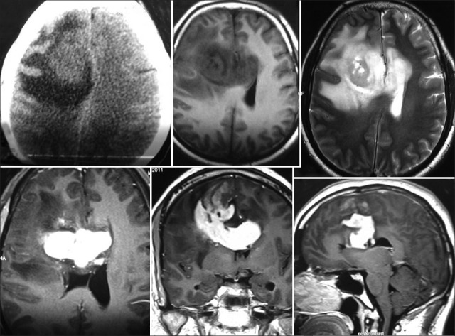
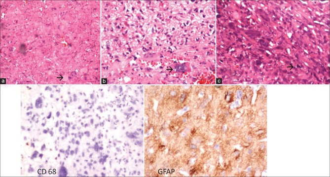
Pleomorphic xanthoastrocytoma (PXA) is an uncommon tumor constitutes less than 1% of all astrocytic glial neoplasms was first reported in 1979. PXA commonly occurs in young patients and manifests itself first as seizures followed by focal neurological deficits. The role of radiotherapy or chemotherapy has not yet been established because of the relative infrequency of this disease. PXA is classified as grade II tumor in the WHO classification of tumors of the CNS. In literature 9 to 20 % PXA may undergo malignant change at recurrence or may display at the time of initial presentation. Malignant transformation is mainly associated with high mitotic activity and necrosis. The criteria for PXA with anaplastic features was five or more mitotic activity per 10 high power fields, necrosis, microvascular proliferation, marked cellular anaplasia, and high Ki-67 labeling indices. PXA with anaplastic features management is highly controversial as very sparse literature is available. We are reporting a case of PXA with anaplastic features with atypical radiology and tried to review the up to date literature regarding this rare tumor.
Keywords: PXA with anaplasia; Pleomorphic xanthoastrocytoma; astrocytoma; management of PXA.
Conflict of interest statement
Figures
References
-
- Giannini C, Scheithauer BW, Burger PC, Brat DJ, Wollan PC, Lach B, et al. Pleomorphic xanthoastrocytoma: What do we really know about it? Cancer. 1999;85:2033–45. - PubMed
-
- Gomez JG, Garcia JH, Colon LE. A variant of cerebral glioma called pleomorphic xanthoastrocytoma: Case report. Neurosurgery. 1985;16:703–6. - PubMed
-
- Kepes JJ, Rubinstein LJ, Eng LF. Pleomorphic xanthoastrocytoma: a distinctive meningocerebral glioma of young subjects with relatively favorable prognosis. A study of 12 cases. Cancer. 1979;44:1839–52. - PubMed
-
- Nakajima T, Kumabe T, Shamoto H, Watanabe M, Suzuki H, Tominaga T. Malignant transformation of pleomorphic xanthoastrocytoma. Acta Neurochir (Wien) 2006;148:67–71. - PubMed
Publication types
LinkOut - more resources
Full Text Sources
Other Literature Sources