. 2015 Dec;1(4):045001.
doi: 10.1088/2057-1976/1/4/045001. Epub 2015 Oct 23.

Noninvasive Surface Imaging of Breast Cancer in Humans using a Hand-held Optical Imager

Affiliations

Noninvasive Surface Imaging of Breast Cancer in Humans using a Hand-held Optical Imager

Sarah J Erickson-Bhatt et al. Biomed Phys Eng Express. 2015 Dec.

Abstract

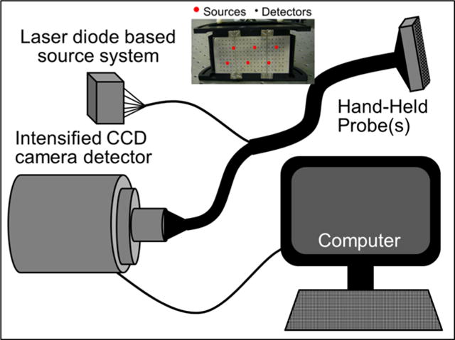
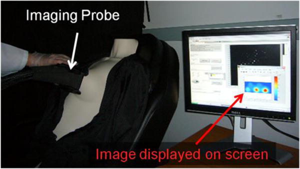
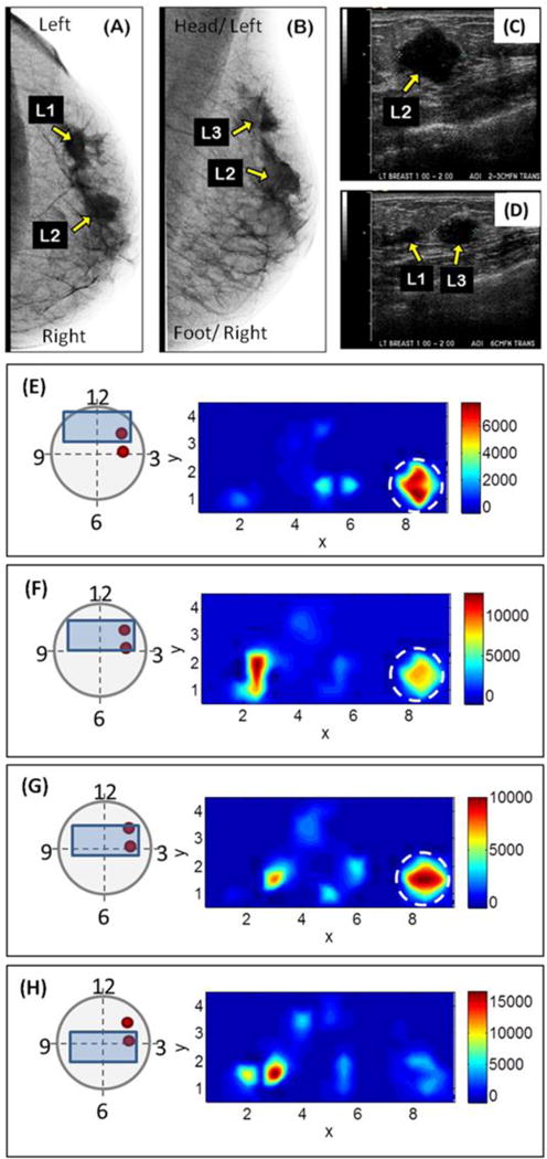
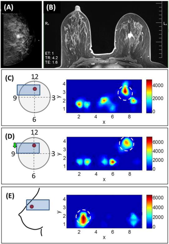
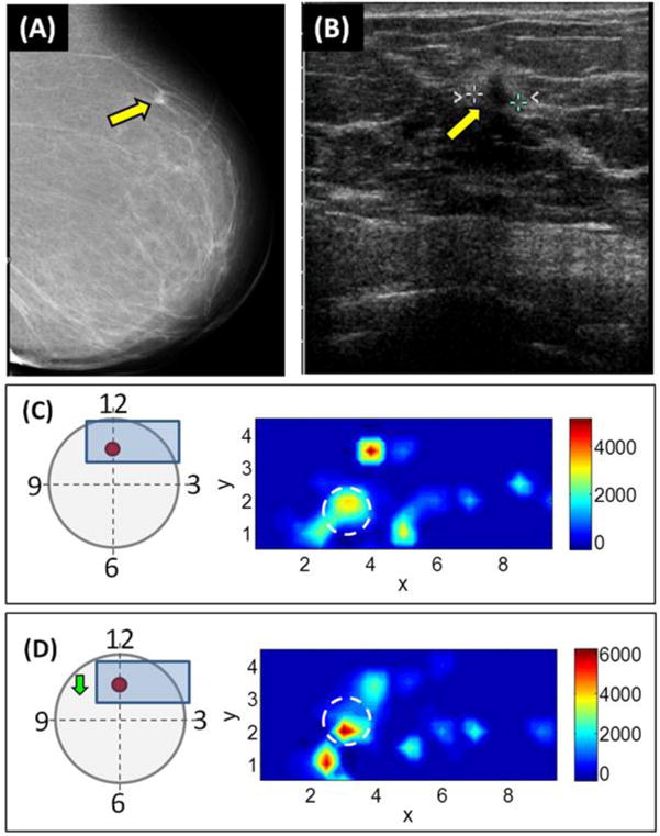
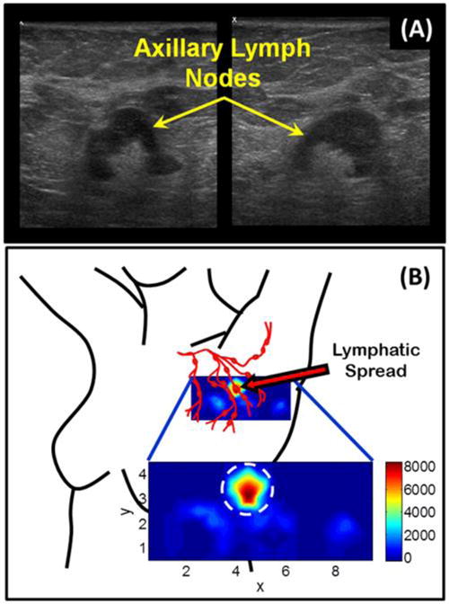
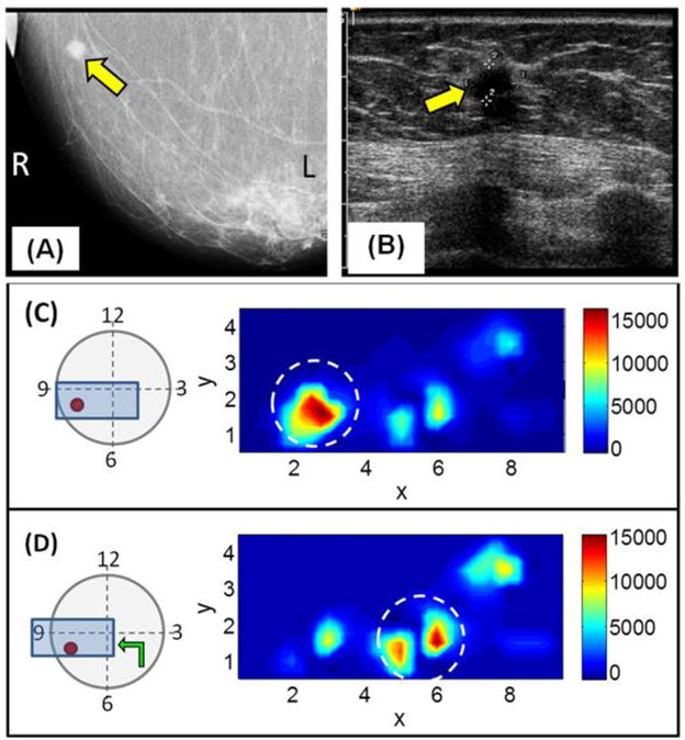
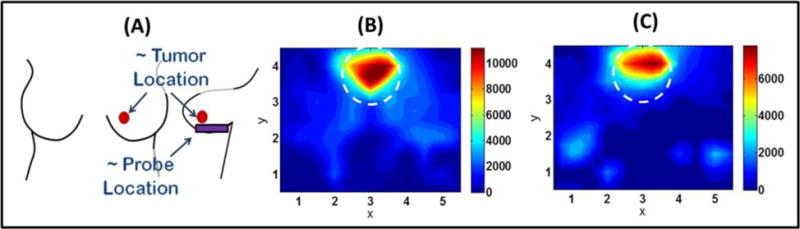
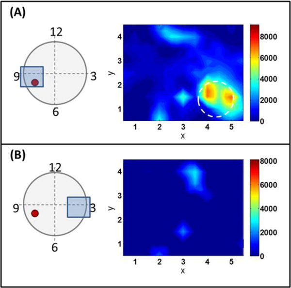
X-ray mammography, the current gold standard for breast cancer detection, has a 20% false-negative rate (cancer is undetected) and increases in younger women with denser breast tissue. Diffuse optical imaging (DOI) is a safe (nonionizing), and relatively inexpensive method for noninvasive imaging of breast cancer in human subjects (including dense breast tissues) by providing physiological information (e.g. oxy- and deoxy- hemoglobin concentration). At the Optical Imaging Laboratory, a hand-held optical imager has been developed which employs a breast contourable probe head to perform simultaneous illumination and detection of large surfaces towards near real-time imaging of human breast cancer. Gen-1 and gen-2 versions of the handheld optical imager have been developed and previously demonstrated imaging in tissue phantoms and healthy human subjects. Herein, the hand-held optical imagers are applied towards in vivo imaging of breast cancer subjects in an attempt to determine the ability of the imager to detect breast tumors. Five female human subjects (ages 51-74) diagnosed with breast cancer were imaged with the gen-1 optical imager prior to surgical intervention. One of the subjects was also imaged with the gen-2 optical imager. Both imagers use 785 nm laser diode sources and ICCD camera detectors to generate 2D surfaces maps of total hemoglobin absorption. The subjects lay in supine position and images were collected at various locations on both the ipsilateral (tumor-containing) and contralateral (non-tumor containing) breasts. The optical images (2D surface maps of optical absorption due to total hemoglobin concentration) show regions of higher intensity at the tumor location, which is indicative of increased vasculature and higher blood content due to the presence of the tumor. Additionally, a preliminary result indicates the potential to image lymphatic spread. This study demonstrates the potential of the hand-held optical devices to noninvasively image breast cancer in human subjects.


Conflict of interest statement

Conflicts of Interest

All authors certify that this manuscript has not been published in whole or in part nor is it being considered for publication elsewhere. The authors have no conflicts of interest to declare.

Figures

Figure 1
Figure 1
Schematic of the CW based hand-held optical imaging system. (Inset) The hand-held probe face (figure is not to scale).
Figure 2
Figure 2
Experimental set-up for in vivo breast imaging.
Figure 3
Figure 3
X-ray, ultrasound, and diffuse optical images of the left breast from a 51 year-old breast cancer patient. (A) X-ray mammography: The top of the image is the left side and the bottom of the image is the right side of the breast tissue. (B) X-ray mammography: Oblique image where the top of the image is toward the patient’s head and left side and the bottom of the image is toward the patient’s feet and right side. The yellow arrows indicate 3 lesions (labeled as L1, L2, and L3) located in the 1 o’clock to 2 o’clock region of the breast. (C–D) Ultrasound images showing two malignant lesions (L1, and L2), and one benign lesion (L3). (E–H) Diffuse optical images collected from the breast tissue of a 51 year-old breast cancer patient. The schematics on the left indicate the approximate location of the tumors (red circles) and the probe (blue boxes) on the breast tissue (figure not to scale). The yellow arrows to the left of the probes in the schematic indicate the movement of the probe between each image and the previous. The white dotted circles in the diffuse optical images to the right indicate a rough estimate of the tumor locations. The red color indicates areas of greater absorption due to higher HbT concentration relative to the background.
Figure 4
Figure 4
(A) X-ray mammography and (B) MRI images from a 52 year-old female with ductal carcinoma in situ. The images show a 1.5 cm diameter mass at the 12 o’clock region of the left breast. (C–E) Diffuse optical images of ductal carcinoma in situ in the left breast of a 52 year-old female. (C–D) Images collected at two probe locations from the front of the breast while the subject lie in supine position. The schematics on the left indicate the approximate location of the tumors (red circles) and the probe (blue boxes) on the breast tissue (figure not to scale). The yellow arrow to the left of the probe in the schematic indicates the movement of the probe between that image and the previous. (E) Image collected from the left side while the subject lie on her right side with her arm raised above her head. The black open circles represent the approximate tumor location. The red signal indicates areas of greater absorption due to higher HbT concentration.
Figure 5
Figure 5
(A) X-ray mammography and (B) ultrasound images from a 74 year-old female with invasive ductal carcinoma (case #3). The yellow arrows indicate the location of a 0.7 cm diameter lesion located at 12 o’clock in the left breast. (C–D) Diffuse optical images from two probe locations on the subject from case #3. The schematics on the left indicate the approximate location of the tumors (red circles) and the probe (blue boxes) on the breast tissue (figure not to scale). The yellow arrows to the left of the probes in the schematic indicate the movement of the probe between each image and the previous. The black open circles indicate the approximate tumor location.
Figure 6
Figure 6
(A) Ultrasound images showing axillary lymph nodes from a subject with metastatic carcinoma in the axillary region of the left breast (case #4). (B) Diffuse optical image of the left axillary region from a female subject with metastatic carcinoma (case #4). The smaller image indicates the location of the probe with respect to the axillary lymph nodes and is zoomed in the larger image. The white dotted circle indicates a region of interest where higher absorption (red) signal corresponding to higher HbT concentration might indicate the lymphatic spread.
Figure 7
Figure 7
(A) X-ray mammography and (B) ultrasound images from a 69 year-old female subject with invasive ductal carcinoma (case #5). The yellow arrows indicate the location of a 0.9 cm diameter tumor at 8 o’clock in the left breast. (C–D) Diffuse optical images collected at two probe locations (indicated by the schematics to the left). The schematics on the left indicate the approximate location of the tumors (red circles) and the probe (blue boxes) on the breast tissue (figure not to scale). The yellow arrow to the left of the probe in the schematic indicates the movement of the probe between that image and the previous. The white dotted circles indicate a region of interest at the approximate tumor location where higher absorption (red signal) corresponding to higher HbT concentration might indicate the increased vasculature (and therefore blood content) due to the presence of the tumor.
Figure 8
Figure 8
(A) Schematic showing the approximate tumor location and probe location while imaging a 69 year-old female subject with invasive ductal carcinoma (case #5). (B–C) Diffuse optical images collected from underneath the breast using the gen-2 imager. The white dotted circles indicate a region of interest at the approximate tumor location where higher absorption (red signal) corresponding to higher HbT concentration might indicate the increased vasculature (and therefore blood content) due to the presence of the tumor.
Figure 9
Figure 9
Diffuse optical images collected with the gen-2 probe at the (A) 9 o’clock and (B) 3 o’clock locations (of the same breast) from a 69 year-old female subject with invasive ductal carcinoma (case #5). Area of higher absorption can be seen at the 9 o’clock position which is over the tumor location, whereas the image from the 3 o’clock location away from the tumor does not show area of high absorption. The white dotted circle indicates a region of interest at the approximate tumor location where higher absorption (red signal) corresponding to higher HbT concentration might indicate the increased vasculature (and therefore blood content) due to the presence of the tumor.

Similar articles

Cited by

References

    1. Breast Cancer Facts and Figures 2011–2012. American Cancer Society; www.cancer.org.
    1. National Cancer Institute Factsheet. www.cancer.gov.
    1. Flexman ML, Kim HK, Gunther JE, Lim EA, Alvarez MC, Desperito E, Kalinsky K, Hershman DL, Hielscher AH. Optical biomarkers for breast cancer derived from dynamic diffuse optical tomography. J Biomed Opt. 2013 Sep 1;18(9):96012. doi: 10.1117/1.JBO.18.9.096012. - DOI - PubMed
    1. Enfield L, Cantanhede G, Douek M, Ramalingam V, Purushotham A, Hebden J, Gibson A. Monitoring the response to neoadjuvant hormone therapy for locally advanced breast cancer using three-dimensional time-resolved optical mammography. J Biomed Opt. 2013 May;18(5):56012. doi: 10.1117/1.JBO.18.5.056012. - DOI - PubMed
    1. Ueda S, Roblyer D, Cerussi A, Durkin A, Leproux A, Santoro Y, Xu S, O’Sullivan TD, Hsiang D, Mehta R, Butler J, Tromberg BJ. Baseline tumor oxygen saturation correlates with a pathologic complete response in breast cancerpatients undergoing neoadjuvant chemotherapy. Cancer Res. 2012 Sep 1;72(17):4318–28. doi: 10.1158/0008-5472.CAN-12-0056. - DOI - PMC - PubMed