

Peripheral retinal non-perfusion and treatment response in branch retinal vein occlusion
- PMID: 27366688
- PMCID: PMC4916143
- DOI: 10.18240/ijo.2016.06.12
Peripheral retinal non-perfusion and treatment response in branch retinal vein occlusion
Abstract
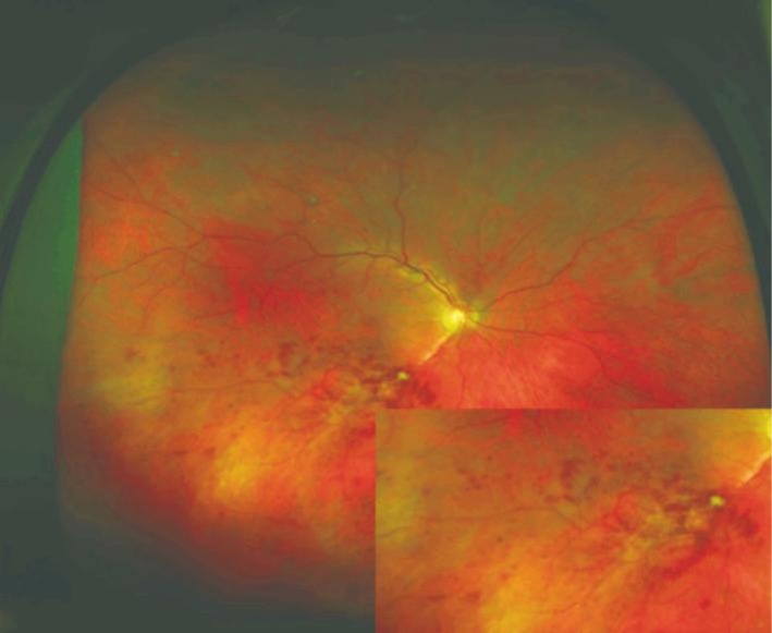
Aim: To evaluate the association between the size of peripheral retinal non-perfusion and the number of intravitreal ranibizumab injections in patients with treatment-naive branch retinal vein occlusion (BRVO) and macular edema.
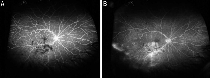
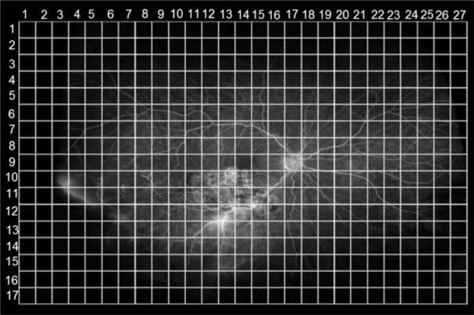
Methods: A total of 53 patients with treatment-naive BRVO and macular edema were included. Each patient underwent a full ophthalmologic examination including optical coherence tomography (OCT) imaging and ultra wide-field fluorescein angiography (UWFA). Monthly intravitreal ranibizumab injections were applied according to the recommendations of the German Ophthalmological Society. Two independent, masked graders quantified the areas of peripheral retinal non-perfusion.
Results: Intravitreal injections improved best-corrected visual acuity (BCVA) significantly from 22.23±16.33 Early Treatment of Diabetic Retinopathy Study (ETDRS) letters to 36.23±15.19 letters (P<0.001), and mean central subfield thickness significantly reduced from 387±115 µm to 321±115 µm (P=0.01). Mean number of intravitreal ranibizumab injections was 3.61±1.56. The size of retinal non-perfusion correlated significantly with the number of intravitreal ranibizumab injections (R=0.724, P<0.001).
Conclusion: Peripheral retinal non-perfusion in patients with BRVO associates significantly with intravitreal ranibizumab injections in patients with BRVO and macular edema.
Keywords: angiography; branch retinal vein occlusion; non-perfusion; retina; wide-field.
Figures
References
-
- Hayreh SS. Prevalent misconceptions about acute retinal vascular occlusive disorders. Prog Retin Eye Res. 2005;24(4):493–519. - PubMed
-
- Ehlers JP, Fekrat S. Retinal vein occlusion: beyond the acute event. Surv Ophthalmol. 2011;56(4):281–299. - PubMed
-
- Rogers SL, McIntosh RL, Lim L, Mitchell P, Cheung N, Kowalski JW, Nguyen HP, Wang JJ, Wong TY. Natural history of branch retinal vein occlusion: an evidence-based systematic review. Ophthalmology. 2010;117(6):1094–1101.e5. - PubMed
-
- Klein R, Moss SE, Meuer SM, Klein BE. The 15-year cumulative incidence of retinal vein occlusion: the Beaver Dam Eye Study. Arch Ophthalmol. 2008;126(4):513–518. - PubMed
LinkOut - more resources
Full Text Sources
Other Literature Sources