. 2016:2016:2414809.
doi: 10.1155/2016/2414809. Epub 2016 Jun 6.

Fibronectin-Grafted Titanium Dental Implants: An In Vivo Study

Affiliations

Fibronectin-Grafted Titanium Dental Implants: An In Vivo Study

Yu-Chi Chang et al. Biomed Res Int. 2016.

Abstract

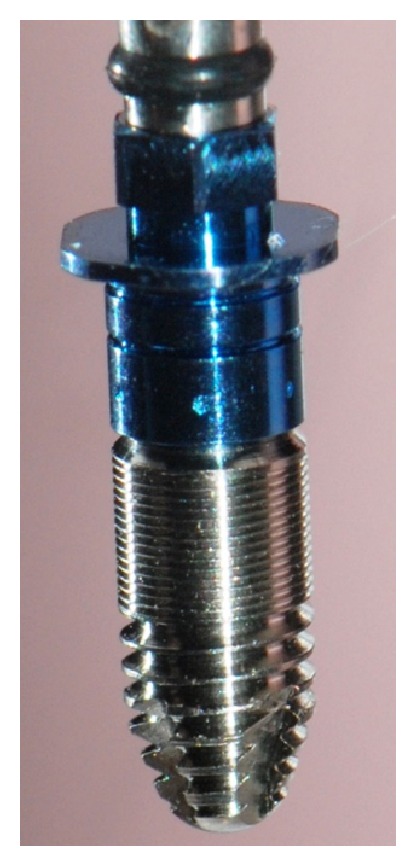
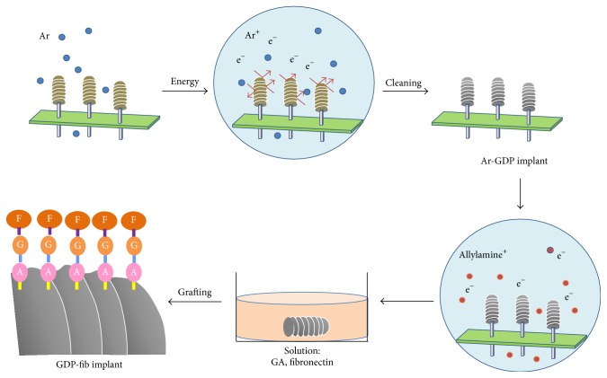
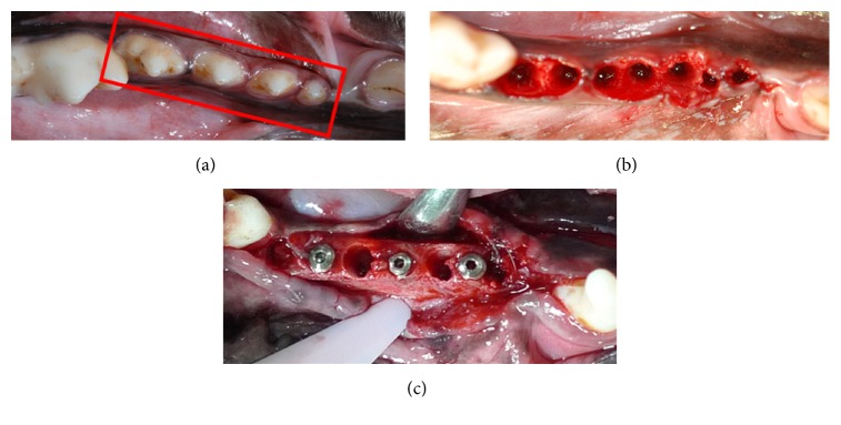
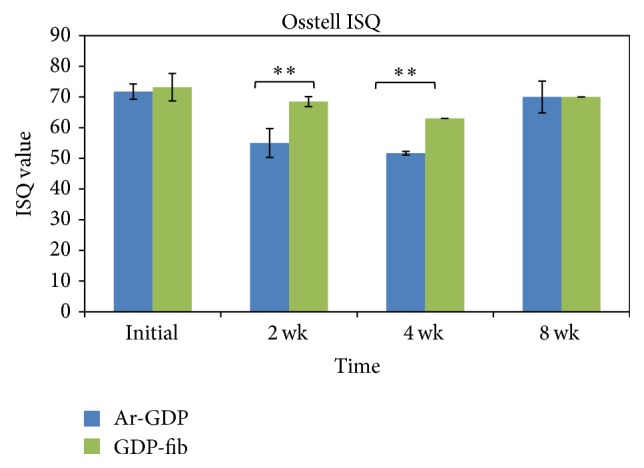
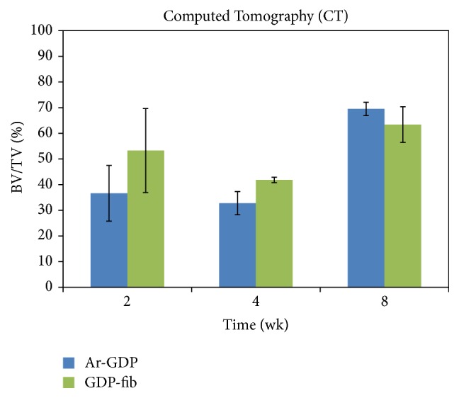
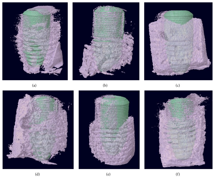
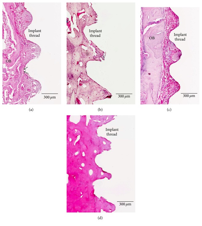
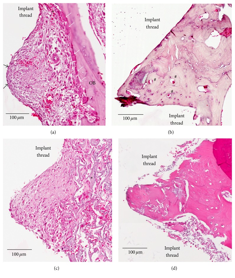
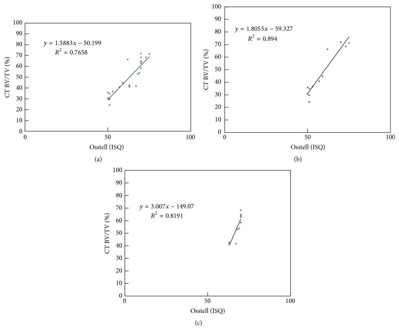
Modification of the physiochemical properties of titanium surfaces using glow discharge plasma (GDP) and fibronectin coating has been shown to enhance the surface hydrophilicity, surface roughness, cell adhesion, migration, and proliferation. This in vivo study aimed to evaluate the bone integration efficacy of a biologically modified implant surface. Two different surface-modified implants (Ar-GDP and GDP-fib) were placed in the mandibular premolar area of six beagle dogs for 2-8 weeks. Three techniques [histologic evaluation, resonance frequency analysis (RFA), and microcomputed tomography (micro-CT) evaluation] were used to detect the implant stability and bone-implant contact. The implant stability quotient values of GDP-fib implants were significantly greater than the Ar-GDP implants at 2 and 4 weeks (P < 0.01). The bone volume/total volume ratio of GDP-fib implants was greater than the Ar-GDP implants in micro-CT evaluation. A high positive correlation was observed between RFA and micro-CT measurements. At 2 weeks, osteoblasts were seen to line the implant surface, and multinuclear osteoclasts could be seen on the surface of old parent bone. After 8 weeks, a majority of the space in the wound chamber appeared to be replaced by bone. Enhancement of the stability of biologically modified implants was proved by the results of RFA, micro-CT, and histological analysis. This enhanced stability may help fasten treatment and be clinically beneficial.


Figures

Figure 1
Figure 1
Experimental implant device: screw-shaped titanium implant packaged with fixture mount. The diameter and length of implant were 3.75 mm and 10 mm, respectively.
Figure 2
Figure 2
Schematic of sample preparation.
Figure 3
Figure 3
(a) Surgical area before extraction. First, second, third, and fourth premolars in the red box. (b) Surgical area after extraction. (c) Placement of implants.
Figure 4
Figure 4
Mean implant stability (implant stability quotient (ISQ) ± SD) of Ar-GDP and GDP-fib implants 2, 4, and 8 weeks after placement. The stability of Ar-GDP implants was significantly lower at 2 and 4 weeks compared to GDP-fib implants (∗∗ P < 0.01).
Figure 5
Figure 5
Mean values of bone-implant contact calculated by micro-CT analysis.
Figure 6
Figure 6
Schematic of Ar-GDP/GDP-fib implants with surrounding bone at 2, 4, and 8 weeks after implantation. (a) Ar-GDP implant at 2 weeks, (b) Ar-GDP implant at 4 weeks, (c) Ar-GDP implant at 8 weeks, (d) GDP-fib implant at 2 weeks, (e) GDP-fib implant at 4 weeks, and (f) GDP-fib implant at 8 weeks.
Figure 7
Figure 7
Ground sections of the peri-implant tissue in the GDP-fib and Ar-GDP groups after 2 and 8 weeks of healing. (a) Wound chamber of the GDP-fib implant at 2 weeks, decalcified section, original mag. ×80. (b) Wound chamber of the GDP-fib implant at 8 weeks, decalcified section, original mag. ×80. (c) Wound chamber of the Ar-GDP implant at 2 weeks, decalcified section, original mag. ×80. (d) Wound chamber of the Ar-GDP device at 8 weeks, decalcified section, original mag. ×80.
Figure 8
Figure 8
Ground sections of the peri-implant tissue of GDP-fib and Ar-GDP implant surfaces after 2 and 8 weeks of healing. (a) Wound chamber of the GDP-fib implant at 2 weeks. Osteoclasts (white arrows) and osteoblasts (black arrows) were surrounded by provisional matrix, decalcified section, original mag. ×200. (b) Wound chamber of the GDP-fib implant at 8 weeks. Osteon (#) could be clearly identified, decalcified section, original mag. ×200. (c) Wound chamber of the Ar-GDP implant at 2 weeks. Woven bone () formation extending into provisional connective tissue matrix was seen, decalcified section, original mag. ×200. (d) Wound chamber of the Ar-GDP implant at 8 weeks, decalcified section, original mag. ×200.
Figure 9
Figure 9
Relationship of the 3D bone-implant contact (BIC) measurements between the BV/TV ratio from CT analysis and the RF data in (a) all, (b) Ar-GDP, and (c) GDP-fib implants.

References

    1. Branemark P.-I. Osseointegration and its experimental background. The Journal of Prosthetic Dentistry. 1983;50(3):399–410. doi: 10.1016/S0022-3913(83)80101-2. - DOI - PubMed
    1. Shalabi M. M., Wolke J. G., Jansen J. A. The effects of implant surface roughness and surgical technique on implant fixation in an in vitro model. Clinical Oral Implants Research. 2006;17(2):172–178. doi: 10.1111/j.1600-0501.2005.01202.x. - DOI - PubMed
    1. Blanco J., Alvarez E., Muñoz F., Liñares A., Cantalapiedra A. Influence on early osseointegration of dental implants installed with two different drilling protocols: a histomorphometric study in rabbit. Clinical Oral Implants Research. 2011;22(1):92–99. doi: 10.1111/j.1600-0501.2010.02009.x. - DOI - PubMed
    1. Rasmusson L., Meredith N., Cho I. H., Sennerby L. The influence of simultaneous versus delayed placement on the stability of titanium implants in onlay bone grafts. A histologic and biomechanic study in the rabbit. International Journal of Oral and Maxillofacial Surgery. 1999;28(3):224–231. doi: 10.1016/s0901-5027(99)80143-x. - DOI - PubMed
    1. Glauser R., Sennerby L., Meredith N., et al. Resonance frequency analysis of implants subjected to immediate or early functional occlusal loading. Successful vs. failing implants. Clinical Oral Implants Research. 2004;15(4):428–434. doi: 10.1111/j.1600-0501.2004.01036.x. - DOI - PubMed

LinkOut - more resources