

Analysis of homo- and hetero-dimerization among the three 6-phosphogluconate dehydrogenase isoforms of Arabidopsis
- PMID: 27366940
- PMCID: PMC5117088
- DOI: 10.1080/15592324.2016.1207034
Analysis of homo- and hetero-dimerization among the three 6-phosphogluconate dehydrogenase isoforms of Arabidopsis
Abstract
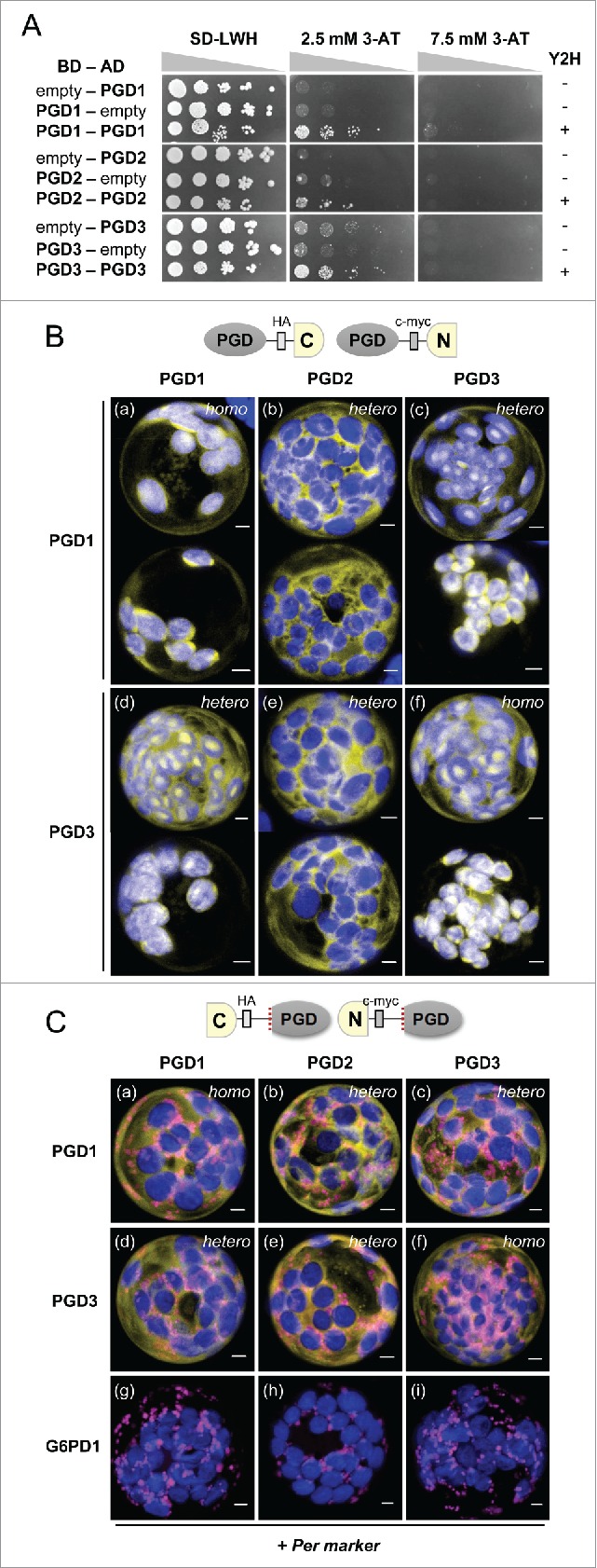
We recently described that all three 6-phosphogluconate dehydrogenase (6PGDH) isoforms of Arabidopsis (PGD) with similar length show dual localization: PGD1 and PGD3 in the cytosol and in plastids, and PGD2 in the cytosol and in peroxisomes. We set out to investigate heterodimer formation, however due to only weak homodimerization of all Arabidopsis PGD isoforms in yeast cells, we conducted further protein-protein interaction studies in planta to investigate homomer versus heteromer formation and their sub-cellular localization. Bimolecular fluorescence complementation (BiFC) analyses in co-transfected Arabidopsis protoplasts demonstrated that all PGD isoforms may form homo- and heterodimers. Notably, with free N-terminal ends, PGD1-PGD3 heterodimers were detected both in the cytosol and in the plastid stroma, but heterodimers with PGD2 only in the cytosol. This independently confirmed that PGD2 cannot enter plastids. On the other hand, with free C-terminal ends, PGD1-PGD2 and PGD3-PGD2 heterodimers were confined to the cytosol, indicating that only PGD2 homodimers are imported by peroxisomes. Together these findings suggest functional redundancy of PGD1 and PGD3 inside plastids, and relevance of PGD1-PGD2 or PGD3-PGD2 heterodimer formation in the cytosol: this could retain sufficient 6PGDH activity needed for NADPH provision, especially during stress defense and initiation of developmental responses.
Keywords: A. thaliana; OPPP; PGD isoforms; dimerization; sub-cellular targeting.
Figures
Similar articles
-
Evidence for dual targeting control of Arabidopsis 6-phosphogluconate dehydrogenase isoforms by N-terminal phosphorylation.J Exp Bot. 2024 May 20;75(10):2848-2866. doi: 10.1093/jxb/erae077. J Exp Bot. 2024. PMID: 38412416 Free PMC article.
-
Defects in Peroxisomal 6-Phosphogluconate Dehydrogenase Isoform PGD2 Prevent Gametophytic Interaction in Arabidopsis thaliana.Plant Physiol. 2016 May;171(1):192-205. doi: 10.1104/pp.15.01301. Epub 2016 Mar 3. Plant Physiol. 2016. PMID: 26941195 Free PMC article.
-
Alternative targeting of Arabidopsis plastidic glucose-6-phosphate dehydrogenase G6PD1 involves cysteine-dependent interaction with G6PD4 in the cytosol.Plant J. 2011 Jun;66(5):745-58. doi: 10.1111/j.1365-313X.2011.04535.x. Epub 2011 Mar 21. Plant J. 2011. PMID: 21309870
-
Evidence for peroxisomal redundancy among the glucose-6-phosphate dehydrogenase isoforms of Arabidopsis thaliana.Plant Cell Physiol. 2025 May 30;66(5):722-737. doi: 10.1093/pcp/pcaf012. Plant Cell Physiol. 2025. PMID: 39829315 Free PMC article.
-
Role and regulation of plastid sigma factors and their functional interactors during chloroplast transcription - recent lessons from Arabidopsis thaliana.Eur J Cell Biol. 2010 Dec;89(12):940-6. doi: 10.1016/j.ejcb.2010.06.016. Epub 2010 Aug 10. Eur J Cell Biol. 2010. PMID: 20701995 Review.
Cited by
-
Evidence for dual targeting control of Arabidopsis 6-phosphogluconate dehydrogenase isoforms by N-terminal phosphorylation.J Exp Bot. 2024 May 20;75(10):2848-2866. doi: 10.1093/jxb/erae077. J Exp Bot. 2024. PMID: 38412416 Free PMC article.
-
6-Phosphogluconate dehydrogenase 2 bridges the OPP and shikimate pathways to enhance aromatic amino acid production in plants.Sci China Life Sci. 2024 Nov;67(11):2488-2498. doi: 10.1007/s11427-024-2567-4. Epub 2024 Jul 24. Sci China Life Sci. 2024. PMID: 39060614
-
Peroxisomes as redox-signaling nodes in intracellular communication and stress responses.Plant Physiol. 2021 May 27;186(1):22-35. doi: 10.1093/plphys/kiab060. Plant Physiol. 2021. PMID: 33587125 Free PMC article. Review.
-
The plastidial pentose phosphate pathway is essential for postglobular embryo development in Arabidopsis.Proc Natl Acad Sci U S A. 2019 Jul 23;116(30):15297-15306. doi: 10.1073/pnas.1908556116. Epub 2019 Jul 11. Proc Natl Acad Sci U S A. 2019. PMID: 31296566 Free PMC article.
References
-
- Kruger NJ, von Schaewen A. The oxidative pentose phosphate pathway: structure and organisation. Curr Opin Plant Biol 2003; 6:236-46; PMID:12753973; http://dx.doi.org/10.1016/S1369-5266(03)00039-6 - DOI - PubMed
-
- Meyer T, Hölscher C, Schwöppe C, von Schaewen A. Alternative targeting of Arabidopsis plastidic glucose-6-phosphate dehydrogenase G6PD1 involves cysteine-dependent interaction with G6PD4 in the cytosol. Plant J 2011; 66:745-58; PMID:21309870; http://dx.doi.org/10.1111/j.1365-313X.2011.04535.x - DOI - PubMed
-
- Hölscher C, Meyer T, von Schaewen A. Dual-targeting of Arabidopsis 6-Phosphogluconolactonase 3 (PGL3) to chloroplasts and peroxisomes involves interaction with Trx m2 in the Cytosol. Mol Plant 2014; 7:252-5; PMID:24008768; http://dx.doi.org/10.1093/mp/sst126 - DOI - PubMed
-
- Foyer CH, Noctor G. Redox homeostasis and antioxidant signaling: a metabolic interface between stress perception and physiological responses. Plant Cell 2005; 17:1866-75; PMID:15987996; http://dx.doi.org/10.1105/tpc.105.033589 - DOI - PMC - PubMed
-
- Scharte J, Schön H, Tjaden Z, Weis E, von Schaewen A. Isoenzyme replacement of glucose-6-phosphate dehydrogenase in the cytosol improves stress tolerance in plants. Proc Natl Acad Sci USA 2009; 106:8061-6; PMID:19416911; http://dx.doi.org/10.1073/pnas.0812902106 - DOI - PMC - PubMed
Publication types
MeSH terms
Substances
LinkOut - more resources
Full Text Sources
Other Literature Sources
Molecular Biology Databases
Miscellaneous