

ASC contributes to metastasis of oral cavity squamous cell carcinoma
- PMID: 27367024
- PMCID: PMC5226569
- DOI: 10.18632/oncotarget.10317
ASC contributes to metastasis of oral cavity squamous cell carcinoma
Abstract
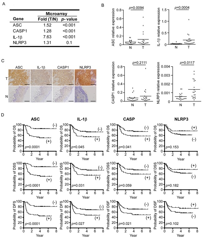
ASC (Apoptosis-associated Speck-like protein containing a CARD) acts as a platform protein in the inflammasome cascade of some cancer types. However, its potential involvement in OSCC (oral cavity squamous cell carcinoma) has not yet been determined. Here, we investigated the potential role of ASC in OSCC. RT-qPCR analysis of 20 paired tumor and adjacent normal tissue samples revealed that the mRNA levels of ASC, along with IL-1β, CASP1, and NLRP3 in ASC-associated NLRP3 inflammasome were significantly elevated in OSCC tissues. Immunohistochemical staining of these four proteins in 111 clinical specimens revealed that high-level expression of ASC was significantly associated with tumor stage, node stage (p=0.001), overall stage (p<0.001), extracapsular spread (p<0.001), perineural invasion (p=0.004) and tumor depth (p<0.001). Kaplan-Meier survival analysis further revealed that high-level ASC expression was correlated with poorer overall survival (p=0.001), disease-specific survival (p<0.001) and disease-free survival (p<0.001). Studies using OSCC cell lines indicated that high-level ASC expression enhanced cell migration and invasion, and experiments using an orthotropic nude mouse model confirmed that ASC overexpression induced metastasis of OSCC cells. This is the first report to show that ASC contributes to OSCC metastasis, and that high-level ASC expression is a marker for poor prognosis in OSCC patients.
Keywords: ASC; OSCC; metastasis.
Conflict of interest statement
The authors report no potential conflict of interest.
Figures


References
-
- Parkin DM, Bray F, Ferlay J, Pisani P. Global cancer statistics, 2002. CA Cancer J Clin. 2005;55:74–108. - PubMed
-
- Reid BC, Winn DM, Morse DE, Pendrys DG. Head and neck in situ carcinoma: incidence, trends, and survival. Oral oncology. 2000;36:414–420. - PubMed
-
- Funk GF, Karnell LH, Robinson RA, Zhen WK, Trask DK, Hoffman HT. Presentation, treatment, and outcome of oral cavity cancer: a National Cancer Data Base report. Head & neck. 2002;24:165–180. - PubMed
-
- Diaz EM, Jr, Holsinger FC, Zuniga ER, Roberts DB, Sorensen DM. Squamous cell carcinoma of the buccal mucosa: one institution's experience with 119 previously untreated patients. Head & neck. 2003;25:267–273. - PubMed
-
- Muir C, Weiland L. Upper aerodigestive tract cancers. Cancer. 1995;75:147–153. - PubMed
MeSH terms
Substances
LinkOut - more resources
Full Text Sources
Other Literature Sources
Medical
Miscellaneous
